It’s wintertime, and the uptick in winter-related illnesses is all around us. The majority of illnesses that tend to peak in the wintertime are caused by viruses, including COVID-19, influenza, and the common cold. Although viruses are generally associated with causing diseases, they may also be an important tool for treating them.
Dr. Jason Kaelber, Associate Research Professor at the Institute for Quantitative Biomechanics at Rutgers New Brunswick in New Brunswick, NJ, is a virologist1 who studies viruses to improve virus-based treatment options for patients. One day, his lab received a package in the mail labeled “live bait.” Dr. Kaelber had no idea why this package was in the mailroom, but he knew who would—a post-doctorate researcher in his lab who specializes in insect viruses, Dr. Judit Pénzes.
Inside that box of live bait lay the clues that would help Dr. Kaelber and Dr. Pénzes identify the cause of a “superworm apocalypse” sweeping across US insect farms and demonstrate the diagnostic power of cryogenic electron microscopy (cryo-EM).
A Box of Bait
Insect farming is a growing industry across the United States and around the world. Insect farmers breed and raise a variety of insects to be used primarily as animal feed for poultry, fish, and pets. The most popular insects for farming include crickets, mealworms, and superworms.
Mealworms and superworms are not real worms. They are the larval stage of certain species of darkling beetles. Mealworms are the larvae of Tenebrio molitor, a darkling beetle commonly known as the yellow mealworm beetle.
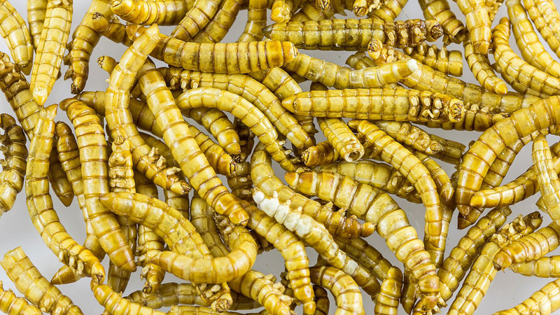
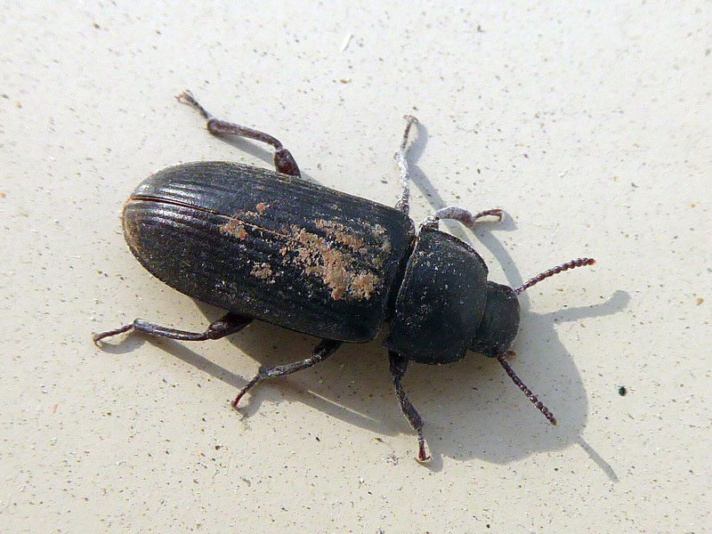
Figure 1.
The yellow darkling beetle Tenebrio molitor in its larval stage, known as a mealworm (top) and as a mature adult (bottom).
[Source: https://commons.wikimedia.org/wiki/File:Mealworms_as_food-2389.jpg; https://commons.wikimedia.org/wiki/File:Tenebrio_molitor_Klaip%C4%97da_01.jpg]
Superworms are the larvae of Zophobas morio. Z. morio larvae can reach up to two inches long and are some of the largest grown as feed, which is why they are commonly referred to as “superworms.”
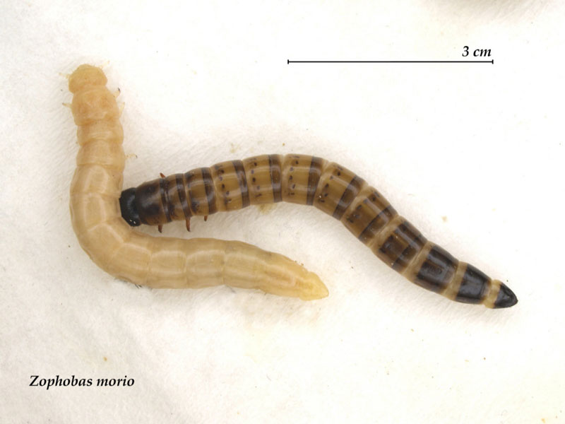
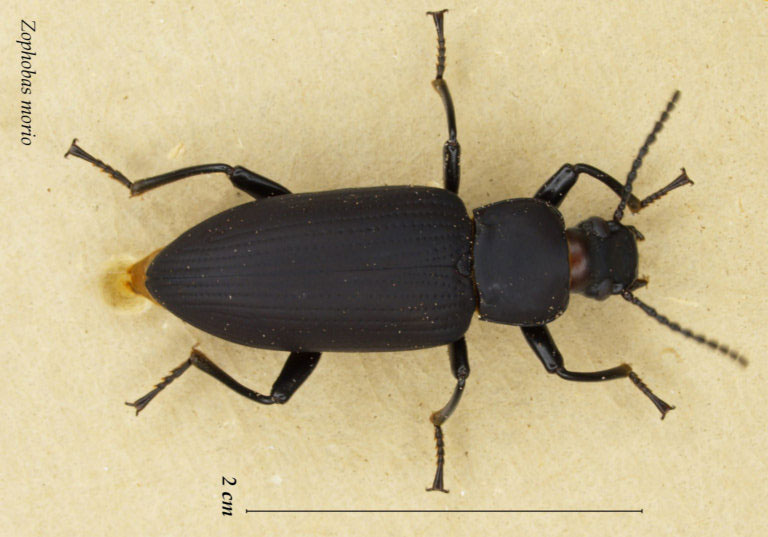
Figure 2.
The darkling beetle Zophobas morio in its larval stage, known as a superworm (top) and as a mature adult (bottom).
[Source: https://commons.wikimedia.org/wiki/File:Zophobas_morio_larvensmall.jpg; https://commons.wikimedia.org/wiki/File:Zophobas_morio_adultsmall.jpg]
Like humans and most other living organisms, insects can be affected by diseases, and diseases that spread rapidly across insect colonies are an important concern for insect farmers. Dr. Pénzes had connections with some of these farmers because of her previous research into cricket viruses2 conducted before joining Dr. Kaelber’s laboratory. As part of this research, farmers would send Dr. Pénzes shipments of abnormal insects that she would screen for viruses and otherwise assist in figuring out the cause of the problem.
Dr. Pénzes first found out about what US farmers were calling a “superworm apocalypse” because she was tagged in a chat forum asking for her help. The requestor was a commercial farmer who had been running a live bait business for over 20 years. “His entire colony of superworms had just died and he wanted answers about what was going on,” recalled Dr. Pénzes. “He offered to ship the entire colony of dead superworms for me to analyze.” These dead superworms were in the package labeled “live bait” that Dr. Kaelber had received.
Superworms affected by the disease would start to show uncoordinated muscle contractions and begin to darken and die.
Diagnosing an Unknown Disease
When Dr. Pénzes first opened the box of dead superworms, she wasn’t sure what to do. Her previous research had focused on cricket viruses, but there were no viruses known to affect superworms. The cause of the disease affecting the superworms could be a fungus3, an environmental issue like humidity, or a virus. Given that the disease killed nearly the entire colony, Dr. Pénzes started by assuming the cause was a virus.
To test whether the superworms were killed by a virus, Dr. Pénzes took the dead superworms and blended them together into a liquid. This liquid was processed using a method called sucrose gradient fractionation that separates molecules in the liquid based on density. The result confirmed that the pathogen causing the disease was a virus, but Dr. Pénzes still didn’t know which one.
Viruses are tiny microbes that can infect the cells of humans, animals, plants, and other organisms. They are too small to be seen with a conventional light microscope4, so researchers have developed various techniques to detect them. Currently, one of the leading methods for viral detection is next generation sequencing5 (NGS). NGS is a complex process that allows researchers to identify the sequence of genetic material in a sample.
The general workflow of NGS involves four basic steps:
- Genetic material needs to be extracted from the sample.
- A library of fragments of the genetic material is created based on what was found in the sample.
- The genetic material of the sample is sequenced, meaning that each individual nucleotide6 is identified.
- The sequencing data is analyzed for relevant results.
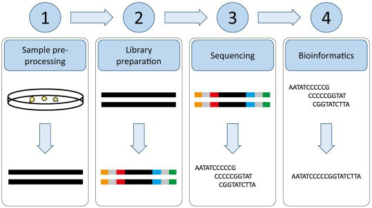
Figure 4.
Overview of the workflow of next generation sequencing or NGS.
[Source: Hess et al 2020, Fig 1]
There are multiple approaches to NGS that vary based on the kind of sample and the type of information researchers are looking for. Like all technologies, NGS has some limitations. It can be particularly challenging to use in research about viruses because viral genetic material varies greatly in size and shape, making it difficult for researchers to synthesize the results.
Another technology for identifying viruses is electron microscopy. Electron microscopy refers to a technique using beams of electrons instead of light to create more detailed images of a sample. Researchers have had some success identifying viruses using electron microscopy by making guesses based on the shape of the resulting image.
Dr. Pénzes and Dr. Kaelber thought that cryogenic electron microscopy7, or cryo-EM, might be better at identifying viruses than traditional electron microscopy or NGS because cryo-EM is detailed enough to show the atoms of the virus in 3D. In cryo-EM, samples are flash-frozen into thin sheets before imaging, making cryo-EM more powerful than conventional electron microscopy. The electron microscope creates a two-dimensional projection of the frozen sample that forms the basis of three-dimensional models. Cryo-EM models have a resolution of 0.2 nanometers. For reference, a human hair is about 80,000-100,000 nanometers wide, while a virus is about 100 nanometers wide. As a result, cryo-EM allows researchers to see the atoms of the virus.
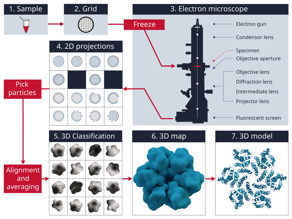
Figure 5.
General workflow when using cryo-electron microscopy (cryo-EM). Cryo-EM allows for the creation of three-dimensional models based on two-dimensional imaging.
[Source: https://commons.wikimedia.org/wiki/File:Cryogenic_electron_microscopy_workflow.svg]
Dr. Kaelber specializes in the use of cryo-EM technology to solve problems in the study of viruses. Since he was already familiar with this technique, Dr. Kaelber and Dr. Pénzes used cryo-EM to analyze the virus sample from the superworms.
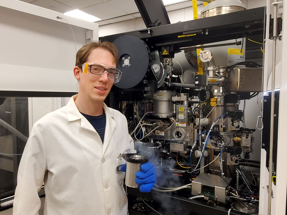
Figure 6.
Dr. Kaelber using the cryogenic electron microscope.
[Source: Dr. Kaelber]
Based on the three-dimensional images from cryo-EM, the researchers constructed a model of the viral structure including its genetic material and proteins. Analyzing this model, they identified a feature of the protein inside the virus that is common to a family of viruses called parvoviruses.
Next, the researchers searched a protein database for similar structures and returned three results—all parvoviruses. This was later confirmed using NGS, which found that the superworm virus was most similar to a virus that affects cockroaches. The researchers named this novel virus Z. morio black wasting virus (ZmBWV). This was the first time that cryo-EM technology had been used as a diagnostic tool to identify an unknown virus.
Proving Cause and Effect
To prove that ZmBWV was the cause of the disease the farmers had seen in the superworms, Dr. Kaelber and Dr. Pénzes relied on four criteria known as Koch’s postulates. If all four of these postulates are met, it demonstrates that a given pathogen causes a disease and is not just correlated with it. The four postulates are:
- A specific microorganism is always associated with a given disease.
- The microorganism can be isolated from the diseased animal and grown in pure culture in the laboratory.
- The cultured microbe will cause disease when transferred to a healthy animal.
- The same type of microorganism can be isolated from the newly infected animal.
In the case of ZmBWV, all four postulates were met:
- The researchers identified ZmBWV in all superworm colonies that showed symptoms of the virus.
- They isolated ZmBWV and grew it in the laboratory by infecting insect cells grown in a dish.
- When uninfected superworms were exposed to the ZmBWV grown in the laboratory, they got the disease.
- ZmBWV was isolated from these uninfected superworms who were exposed to ZmBWV and subsequently got the disease.
By meeting all four of Koch’s postulates, Dr. Kaelber and Dr. Pénzes were able to definitively prove that ZmBWV was the cause of the disease farmers had noticed affecting their superworm colonies.
As part of her collaboration with insect farmers, Dr. Pénzes shared this information with the community. Additional farmers saw Dr. Pénzes’s work and sent more samples to Dr. Kaelber’s laboratory. Analyzing these samples, Dr. Kaelber and Dr. Pénzes realized that the same strain of parvovirus was infecting superworm colonies all over the United States.
Origins of a Virus
At this point, Dr. Pénzes still didn’t know where the parvovirus infecting superworms might have come from. To investigate, she observed the insect bins at different pet stores. At one pet store, she saw that some of the mealworms were diseased and looked much like the dead superworms. With the permission of the pet store owner, she took samples of both dead mealworms and live ones. After purification with sucrose gradient fractionation and imaging with cryo-EM, Dr. Pénzes found that all the mealworms—both dead and alive—had the same virus found in the superworms. The virus was affecting the mealworms but not causing the devastation seen in superworms. “We concluded that there must be two strains of this parvovirus, one that causes disease and one that doesn’t,” commented Dr. Pénzes.
Since there were two strains of the disease, Dr. Pénzes hypothesized that she could use the viral strain that doesn’t cause disease to vaccinate the superworms against the strain that does. “It is important to note that vaccination in an agricultural context is not the same as in human medicine,” added Dr. Kaelber. Unlike human medicine, vaccines for agriculture need to be low cost and low tech—and not every insect needs to survive.
Dr. Kaelber and Dr. Pénzes are in the process of optimizing the ZmBWV vaccine to be most effective and easy to deliver. The current vaccination strategy is to deliver the vaccination with a viral strain that doesn’t cause disease in Jello-like sheets that the superworms would eat.
Future Work
Dr. Kaelber and Dr. Pénzes are taking this work in multiple directions. Dr. Pénzes will soon be starting a new position as an Assistant Professor of Entomology8 at Texas A&M University where she will continue to work on improving our understanding of insect viruses. “Insects are the most populous type of species in the animal kingdom,” explained Dr. Pénzes, “and we know so little about them.” The parvovirus that infected superworms was the first known virus in beetles. “We are just scratching the surface,” she added.
Dr. Kaelber is excited about the insights from insect viruses that might transform human medicine. His research focuses on designing viruses that could be useful for humans. “Viruses don’t just cause disease,” explained Dr. Kaelber, “they are also an effective tool for delivering treatment.” One example of this is gene therapy9, which seeks to alter our genetic material to help us fight disease. The gene therapy must be carried to cells via a vector10, and viral vectors are a common strategy. “We’re hoping to use properties of insect viruses that we identify to design more effective viruses,” Dr. Kaelber added.
In addition, Dr. Kaelber hopes to contribute to advancing the technology of cryo-EM so it can aid in more powerful tasks. The parvovirus in superworms is the first time that cryo-EM has been used as a diagnostic tool to identify an unknown pathogen.
Dr. Jason Kaelber is Associate Research Professor at the Institute for Quantitative Biomechanics at Rutgers New Brunswick and Director of the Rutgers CryoEM & Nanoimaging Facility. His research focuses on using cryo-electron microscopy to solve challenging biological problems such as virus design for human medicine. Outside of the lab, Dr. Kaelber co-leads a homeless shelter in New Brunswick, New Jersey where he works with homeless guests and volunteers. “In science and research, you might not see the impact of your work for decades to come,” commented Dr. Kaelber. “I enjoy volunteering and seeing the immediate impact of our work.”
Dr. Judit Pénzes is a post-doctoral researcher in Dr. Kaelber’s lab who specializes in insect viruses. In addition to her research, Dr. Pénzes is part of the World Health Organization’s Pandemic Prevention Initiative. This initiative seeks to shortlist certain pathogens so that scientists and clinicians are more prepared in the case of a future pandemic. There is a study group for each family of viruses, and Dr. Pénzes participates in the parvovirus study group. Outside of the lab, Dr. Pénzes volunteers by re-homing retired greyhounds. She is also an accomplished artist.
For More Information:
- Pénzes JJ, Holm M, Yost SA, Kaelber JT. Cryo-EM-based discovery of a pathogenic parvovirus causing epidemic mortality by black wasting disease in farmed beetles. Cell. 2024;187(20):5604-5619.e14. doi:10.1016/j.cell.2024.07.053
- Pénzes JJ, Pham HT, Chipman P, Smith EW, McKenna R, Tijssen P. Bipartite genome and structural organization of the parvovirus Acheta domesticus segmented densovirus. Nat Commun. 2023;14(1):3515. Published 2023 Jun 14. doi:10.1038/s41467-023-38875-x
To Learn More:
Cryo-Electron Microscopy
- Kaelber Laboratory. https://molbiosci.rutgers.edu/faculty-research/faculty/faculty-detail/84-k-l/1093-kaelber-jason
- Rutgers CryoEM & Nanoimaging Facility. https://iqb.rutgers.edu/RCNF
- “Getting Started in cryo-EM” by Prof. Grant Jensen https://www.youtube.com/watch?v=q17nGZPCeoA
Viruses
- Viruses. https://www.genome.gov/genetics-glossary/Virus
- Parvoviruses. https://www.ncbi.nlm.nih.gov/books/NBK7715/
Insect Farming
- Global Insect Farming Market. https://market.us/report/insect-farming-market/
- Superworms: The Bigger, Brawnier, Hungrier Cousins of Yellow Mealworms. Entomology Today. https://entomologytoday.org/2021/05/20/superworms-zophobas-morio-bigger-brawnier-hungrier-cousins-yellow-mealworms/
Written by Rebecca Kranz with Andrea Gwosdow, PhD at www.gwosdow.com

