| |
Highlights
- Prolonged stress can be detrimental to your health. Studies of people’s reactions to stress found that some people take a long time to recover from stress while others recover quickly.
- A person’s ability to recover from stress is related to the expression of the gene, miR-29c. The expression of miR-29c was higher in people who recovered from stress more slowly.
- A blood test to measure miR-29c expression is being developed to identify patients who do not recover from stress and may need help.
When was the last time you “stressed out” about something? Exams, upcoming deadlines, who to take to the school dance—all of these can induce a
stress response
in both children and adults. Oftentimes, stress can be a positive force, helping us stay energized and focused. In order for stress to remain a positive condition, however, we must be able to recover quickly.
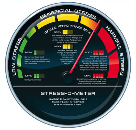
Image from http://fortcollinsbiofeedback.com
Some people are better at this than others, but we are not always the best judges of our own response to stress. New research by Dr. Noam Shomron and Prof. Talma Hendler at Tel Aviv University has identified specific blood markers that can differentiate between those of us who recover more quickly or slowly from stress. The researchers hope that their findings will provide the groundwork for a blood test to help individuals determine, and thereby improve, their response to stress.
It is well documented that prolonged stress can be detrimental to our physical health. See our November 2013 What A Year! article Don’t Stress Out! featuring research by Drs. Marti Jett and Rasha Hammamieh of the U.S. Army Center for Environmental Health Research. Drs. Jett and Hammamieh focused on the ways that extended periods of stress alter
gene expression
of proteins involved in the
immune system,
resulting in abnormal immune function.
It is well documented that prolonged stress can be detrimental to our physical health.
How stress affects our brains
Dr. Shomron and Prof. Hendler have taken a different approach to stress, examining how short periods of stress alter activity in the brain. These researchers are colleagues at Tel Aviv University with distinct areas of expertise. Prof. Hendler specializes in the use of
functional magnetic resonance imaging
(fMRI) to study activity in different areas of the brain. Dr. Shomron uses state-of-the-art genetic sequencing techniques to analyze gene expression. Both researchers work on a variety of topics. For the study of stress, Dr. Shomron and Prof. Hendler joined forces to examine how stress affects activity and gene expression in the brain.
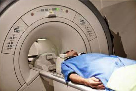
An fMRI machine and patient
The researchers focused on 45 male soldiers aged 19-22 who were assigned to the same combat unit but before operational deployment. Each soldier participated in a standard half-day procedure to collect fMRI data of brain activity and physiological data including blood and saliva samples. Blood samples were taken before and after the MRI scans. Saliva samples and self-evaluations of stress levels were taken at several points before, during, and after the scans.
The fMRI session was divided into five parts: first rest scan, control task scan, second rest scan, stress task scan, and third rest scan. During rest scans, the participants were instructed to stare at a fixed point in the MRI machine. In the control task scan, the participants were told to count backwards from 1,000 and report out loud to the researchers. The participants were given no feedback as to speed or accuracy. This was designed to be a minimally stressful situation. In the stress scan, the participants were told to count backwards from 1,022 by intervals of 13 and report out loud to the researchers. This time, the participants were consistently given feedback to conduct the task more quickly and accurately. The stress scan was designed to be a stressful challenge.
Each soldier participated in a standard half-day procedure to collect fMRI data of brain activity and physiological data including blood and saliva samples.
Once data was obtained from all participants, the researchers analyzed brain activity and gene expression according to their areas of expertise. Prof. Hendler homed in on one area of the brain called the ventro-medial prefrontal cortex (vmPFC) because it is known to be a key area of stress regulation in the brain. Specifically, Prof. Hendler and her team were interested in activity in the vmPFC that involved communication with other parts of the brain. To determine this type of activity, the researchers used a method called Functional Connectivity to analyze the amount of connectivity between the vmPFC and surrounding areas of the brain.
Once brain activity had been quantified for distinct time periods during the scan (rest vs. control task vs. stress task), the researchers compared these results to the levels of stress self-reported by the participants. In doing so, Prof. Hendler and her colleagues found that soldiers fell into two basic groups: those who recovered quickly from stressful events and those who did not. In the soldiers that recovered quickly, activity in the vmPFC increased from baseline and then returned to baseline shortly afterwards. In soldiers that took longer to recover, the brain activity took longer to return to baseline or, in some cases, did not return to baseline by the end of the experiment.
Furthermore, how a soldier said he felt did not necessarily correlate with what was going on physiologically in the brain. Even when a soldier self-reported that he no longer felt stressed, the fMRI scans were sometimes telling a different story.
Blood samples
Meanwhile, Dr. Shomron and colleagues analyzed the blood samples obtained from the participants for differences in gene expression before and after the MRI session. Dr. Shomron suspected that genes with altered expression would be involved in the stress response and might play a role in the differences in brain activity found by Prof. Hendler.
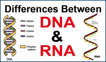
Genetic expression can be measured in a variety of ways. The most basic genetic molecule is
deoxyribonucleic acid
or DNA. Each cell in our bodies contains many copies of DNA. It is a very stable molecule and changes can be observed over long periods of time. Since Dr. Shomron was interested in more subtle changes in genetic expression, DNA was not a good choice for analysis.
http://www.microbiologyinfo.com/differences-between-dna-and-rna/
Instead, Dr. Shomron and his colleagues analyzed changes in molecules of
ribonucleic acid
or RNA. RNA molecules are produced as part of the process that creates proteins based on DNA. They are copies of specific genes, and they are much less stable than DNA. RNA molecules are produced in response to small changes and are therefore a better target for the type of analysis Dr. Shomron conducts.
There are thousands of pieces of RNA floating around in the blood at any given time.
In order to determine how the RNA found in blood samples taken before and after the MRI session differed, Dr. Shomron used machines that are able to read thousands of them in one experiment. In the end, Dr. Shomron identified a handful of genes whose RNA expression was different between the two samples. Of these genes, one in particular was shown to be involved in the stress response. For this reason, Dr. Shomron and colleagues focused on this particular gene called miR-29c. miR-29c is a small molecule of RNA, called
microRNA.
MicroRNAs control gene expression in human cells and are thought to be master regulators of cellular processes.
miR-29c Results
Once Dr. Shomron and Prof. Hendler analyzed their respective data sets, they worked together to combine their results and determine how stress affects both brain activity and genetic expression—and how the two are related. The researchers observed interesting patterns in their data. The soldiers that recovered quickly according to Prof. Hendler’s analysis exhibited no significant changes in miR-29c expression before or after the experiment. However, the difference in expression was remarkable in soldiers that Prof. Hendler identified as recovering more slowly. In these participants, levels of miR-29c were still greatly increased over baseline at the end of the 3-hour experiment.
Based on their results, Prof. Hendler and Dr. Shomron hypothesized that miR-29c could be a useful biological marker for how individuals handle stressful situations. Dr. Shomron envisions a simple blood test that would indicate whether someone recovers more slowly or quickly after a stressful event. This information could then be used on a personal level to help patients better manage their stress.
This is no small matter. As Drs. Jett and Hammamieh showed the inability to recover from stress can cause significant physical problems down the road. The same is true of mental illnesses like
depression
and
post-traumatic stress disorder.
“If we can use this blood test to identify a patient who is not recovering well from a stressful event,” Dr. Shomron explains, “then we can make sure that patient connects with the resources he or she needs.” These resources may include help from a therapist or counselor or general training in how to calm oneself after stress using techniques from meditation, yoga, or other practices.
There are many years of research ahead before this can be done. First, Dr. Shomron plans to use mouse models to further study the effects of miR-29c on brain activity. In addition, Dr. Shomron and Prof. Hendler plan to follow the soldiers participating in this study throughout their training and deployment period. If the results from these experiments support previous findings on miR-29c, the researchers will begin to develop applications such as a blood test. “We still have a long way to go,” Dr. Shomron concludes,” but we are very encouraged by our results and our collaborative efforts.”
Dr. Noam Shomron is Principal Investigator at Sackler Medical School, Tel Aviv University, Tel Aviv, Israel. In his Genomics Intelligence Laboratory, Dr. Shomron analyzes gene expression in a variety of healthy and disease states. When not in the laboratory, Dr. Shomron enjoys spending time with his family.
Prof. Talma Hendler is Director of the Tel Aviv Center for Brain Functions at Tel Aviv University, Tel Aviv, Israel. Her laboratory specializes in the use of functional magnetic resonance imaging.
For More Information:
- Vaisvaser, S. et al. 2016. “Neuro-Epigenetic Indications of Acute Stress Response in Humans: The Case of Micro-RNA-29c. PLoS One.
- Weinglass, Simona. “Is Your Life Too Stressful?” Times of Israel, 25 February 2016.
http://www.timesofisrael.com/is-your-life-too-stressful-there-may-soon-be-a-blood-test-to-find-out/
To Learn More:
- Noam Shomron website. http://nshomron.wix.com/shomronlab
- Tel Aviv Center for Brain Function. http://fmri-tlv.org/
Additional Applications of Dr. Shomron’s work
Written by Rebecca Kranz with Andrea Gwosdow, PhD at www.gwosdow.com
HOME | ABOUT | ARCHIVES | TEACHERS | LINKS | CONTACT
All content on this site is © Massachusetts
Society for Medical Research or others. Please read our copyright
statement — it is important. |
|
|

Dr. Sharon Vaisvaser

Dr. Shira Modai
Dr. Noam Shomron
(photo credit: Sion Ninio)
Prof. Talma Hendler

Dr. Shomron’s TedX talk at Tel Aviv University
Sign Up for our Monthly Announcement!
...or  subscribe to all of our stories! subscribe to all of our stories!

What A Year! is a project of the Massachusetts
Society for Medical Research.
|
|
