| |
Highlights
Acral melanoma is a rare form of melanoma that typically affects the palms of the hands, soles of the feet, and nailbeds. These researchers developed a zebrafish model using the genetic changes seen in human patients with acral melanoma. They found that zebrafish developed tumors predominantly on the fins, the evolutionary equivalent to the hands and feet of people. Further genetic modeling identified overexpression of the gene CRKL as the primary mechanism for the development of acral melanoma. The hands, feet, and fins have increased levels of the proteins HOX13 and insulin-like growth factor (IGF), which work together with CRKL to cause the disease. These results led the researchers to determine positional identity—where a cell is located in the body—as an important factor in determining whether and how cancer might develop. Future work will investigate how positional identity might impact the development and treatment of other types of cancers.
Over ten years ago, a patient with a rare form of melanoma came to Memorial Sloan Kettering Cancer Center (MSKCC) in New York City for treatment. This patient’s case inspired Dr. Richard White, cancer genetics researcher at MSKCC, and his student, Dr. Joshua Weiss, to consider not only a cancer’s genetic profile but also its location in the body as a key factor for cancer development. The researchers hope their results can lead to more targeted forms of cancer treatment in the future.
A Rare Form of Melanoma
Melanoma
is a form of skin cancer that begins in
melanocytes,
specialized skin cells that produce the pigment
melanin.
It is a less common form of skin cancer but can be much more dangerous because it can quickly spread to other parts of the body if not treated early. Melanoma can appear as a new mole, freckle, or as discoloration of the skin (Figure 1).
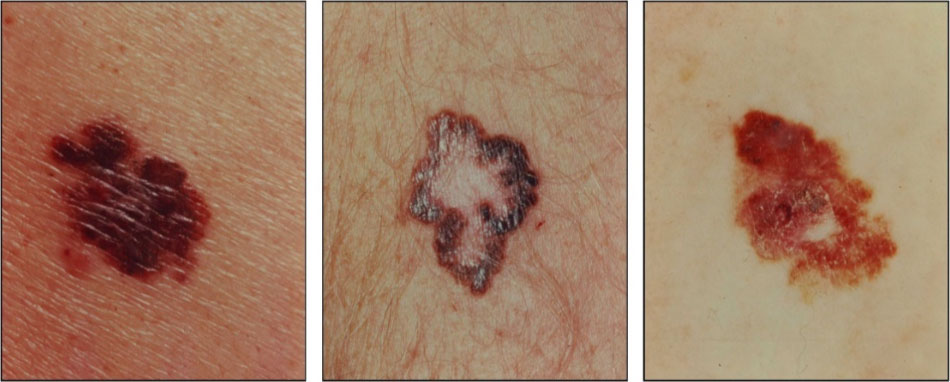
Figure 1. Images of the most common forms of melanoma lesions
[Source: https://nci-media.cancer.gov/pdq/media/images/765750.jpg]
Melanoma does not just affect older adults. According to St. Jude Children’s Research Hospital, melanoma makes up 1% of all cancers in children under 15 years of age and 7% of all cancers in adolescents 15-19 years of age. It most commonly appears on the legs of women and on the trunk or back of men. However, it is important to know that melanoma can appear anywhere on the body.
Early detection is the key to melanoma treatment. Cancer treatment success is often measured by a five-year survival rate, meaning the likelihood that a patient will still be alive five years after diagnosis. According to the American Cancer Society, patients whose melanoma is detected early, before it spreads to other organs, have a five-year survival rate of 99%. However, once melanoma spreads to the
lymph nodes,
the survival rate drops to 68%, and once it has spread to other organs the survival rate drops even lower. This is why it is so important to know the early warning signs of melanoma.
Clinicians and researchers recommend the ABCDE rule for early detection of melanoma:
- A: Asymmetry - most melanomas are asymmetrical, meaning that if you draw a line through the middle, the two sides will look different.
- B: Border - The border or edge of a melanoma tends to be uneven or jagged, whereas noncancerous moles usually have smooth edges.
- C: Color - Multiple colors or shades of brown is a warning sign of melanoma. Noncancerous moles tend to be the same color throughout.
- D: Diameter - Any marking on the skin that grows to be at least the size of a pencil eraser (about ¼ inch in diameter) should be evaluated for melanoma. In addition, any marking on the skin that is darker in color than other markings could also be a warning sign of melanoma.
- E: Evolving - Any changes in a marking, such as its size or shape, or new bleeding or crusting, could be a warning sign for melanoma.
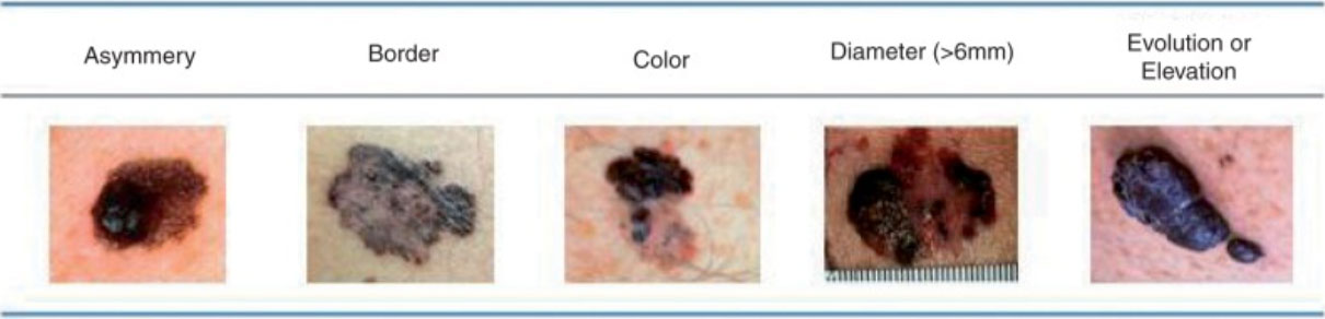
Figure 2. Illustration of the ABCDEs of melanoma
[Source: https://www.ncbi.nlm.nih.gov/books/NBK481857/figure/chapter6.f1/]
There are four main types of skin melanoma:
- Superficial spreading melanoma – this is the most common type of melanoma, which begins by affecting the outermost layers of the skin.
- Nodular melanoma – the second most common type of melanoma, it can be aggressive because it grows quickly. It usually appears as a firm node or bump that rises above the surface of the skin.
- Lentigo maligna melanoma – a type of melanoma that usually affects older people with skin that has been heavily damaged by the sun.
- Acral melanoma - a rare form of melanoma that typically affects only the palms of the hands, soles of the feet, and nailbeds. Acral melanoma tends to respond less well to treatment, leading patients to have less positive outcomes. Researchers know that acral melanoma can often have a different genetic profile compared to other types of melanoma, and it is found in different parts of the body. Why this is the case is not understood.
It is important to note that skin color can be a factor in increasing or decreasing your risk for developing certain kinds of melanoma. Lighter skin color or skin that burns easily can increase the risk of skin cancer in general and some types of melanoma in particular. The most common form of melanoma in people of African and Asian descent is the rarer acral melanoma.
A Zebrafish Model of Acral Melanoma
The patient who inspired ongoing work in Dr. White’s laboratory had acral melanoma, this rare form of melanoma that is poorly understood. A sample of this patient’s cancer tissue was sent to Dr. White’s laboratory for analysis because the White Laboratory specializes in cancer genetics.
The genetic profile of the patient’s cancer did have several altered genes, some of which Drs. White and Weiss had seen in cancer patients previously and some that were new. “From a theoretical perspective, we could understand how these genes might be involved in cancer,” explained Dr. Weiss, “but we hadn’t actually seen a profile like this before.” Specifically, there were four genes that were altered that the researchers investigated further: CRLK (pronounced “crackle”), GAB2, TERT, and NF1.
In order to better understand this patient’s cancer profile, Drs. White and Weiss began conducting experiments in zebrafish, the primary animal model used in Dr. White’s laboratory. Zebrafish are a useful model for understanding genetic changes because their genes are easy to manipulate, allowing researchers to create genetically complex animal models. In other words, rather than making a single genetic change at a time, it is possible to make multiple changes, such as increasing the genetic expression of some genes while removing other genes, all within the same model. In addition, a strain of zebrafish called casper are fairly transparent, and there are a wide variety of imaging tools to visualize their growth and development.
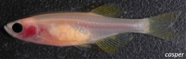
Figure 3. Transparent zebrafish model
[Source: White Lab, https://www.mskcc.org/research/ski/labs/richard-white/postdoctoral-position-modeling-metastasis-zebrafish]
To create a zebrafish model, the first step is to let the zebrafish breed. Zebrafish offspring are very small when they are first born but still visible under the microscope. The researchers take the zebrafish at this stage, when the offspring are only one cell, and inject them with genetic information.
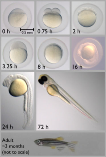
Figure 4. Stages of zebrafish development. Drs. White and Weiss create genetic models of zebrafish by injecting the embryos with genetic information at the single-cell stage.
[Source: https://en.wikipedia.org/wiki/Zebrafish]
The changes the researchers have made are then incorporated into the growing zebrafish, and in one to two months the results can be observed.
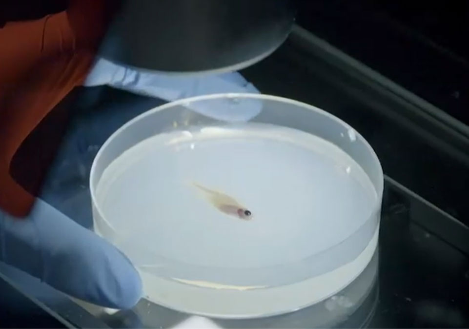
Figure 5. Observing zebrafish under the microscope
[Source: https://www.mskcc.org/videos/look-inside-richard-white-lab-msk]
The first zebrafish model Drs. White and Weiss created in this series of experiments had the exact same genetic expression as the human patient with acral melanoma. The goal was to see how these genetic changes might impact the developing zebrafish. At the time, the researchers were thinking only about this one patient. “We thought this patient’s cancer had a unique genetic profile,” Dr. Weiss commented. However, this impression changed as the researchers began to look back at the genetic profiles of other patients with acral melanoma. “We realized that what we thought was unique was actually a somewhat common genetic profile for this specific type of melanoma,” explained Dr. Weiss.
Just like the human patient with the same genetic profile, the zebrafish model also developed melanoma.
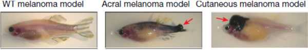
Figure 6. Zebrafish models. Wildtype (WT) or control on the left; acral melanoma in the middle; and cutaneous (superficial spreading) melanoma on the right. The red arrow indicates the location of the melanoma.
[Source: Weiss et al, 2022, Fig 1D]
Moreover, the melanoma tumors that developed seemed to occur mainly on the fins of the zebrafish. These results led the researchers to expand their thinking about what might be happening in acral melanoma to include an evolutionary perspective. This is because, from an evolutionary perspective, “the genes involved in the development of human hands and feet are the same genes involved in fin development in fish,” explained Dr. White.
CRKL Expression in Acral Melanoma
To better understand the effects of each of the four genes altered in the human patient’s genetic profile, Drs. White and Weiss created additional zebrafish models where each model lacked one of the four genes (CRLK, GAB2, TERC, and NF1). This allowed the researchers to individually test the effect of each gene. From this experiment, the researchers identified the gene CRKL as playing a crucial role in the development of melanoma in zebrafish.
The researchers showed that solely by overexpressing the gene CRKL, the zebrafish would start to grow tumors on their fins. The function of the CRKL gene is not well understood. What is known is that it functions as a scaffolding protein by binding to receptor proteins and amplifying a specific signal. CRKL functions differently from most cancer-causing genes, which tend to be altered in such a way that their function is turned permanently on or off. “CRKL itself is normal in the sense that it has no mutations,” added Dr. Weiss, “but it can be amplified or overexpressed in tumors.” The researchers found that the only difference in the CRKL gene in the zebrafish models of melanoma compared to controls is that CRKL is overexpressed. There is more gene than usual, meaning that whatever cancer signals might exist get amplified.
The researchers began to consider what was so special about CRKL, particularly in fish fins and human hands and feet. They found that these areas of the body have increased levels of two groups of molecules called HOX13 and insulin-like growth factor (IGF). “It appears that CRKL works very well with HOX13 and IGF to amplify tumor signals,” explained Dr. White. “We think this might explain why acral melanoma develops only in these specific locations.”
Oncogenetic Competence and Positional Identity
Based on these results, Drs. White and Weiss suggest reframing how cancer develops. “We know that cancer is, in part, a result of genetic mutations,” explained Dr. White. “Our results suggest that, while genetic mutations are a necessary component of cancer development, they are not sufficient.” In other words, there are other factors at play in cancer development aside from genetics. This broad view of why cancer develops in some locations or situations, or in some patients and not others, is what Dr. White calls
oncogenic competence,
meaning the ability of a tissue to become cancerous.
To better understand the concept of oncogenic competence, think about our healthy tissues as a lake. As long as the water in the lake stays below a certain level, the tissue remains healthy and functions properly. Cancer can develop if the water overflows, and there are many reasons why this might happen. For example, if you drop a huge boulder into the middle of the lake. This is what happens in some kinds of cancers where a single genetic mutation can trigger rapid uncontrolled cell growth.
Much of cancer research has focused on these types of genetic mutations. However, most types of cancer are not caused by a single boulder-like event but by smaller pebble-like events that accumulate over time. The overexpression of CRKL is a pebble-like event. In and of itself, it won’t cause acral melanoma. But together with many other factors, it can.
When talking about cancer development, Drs. White and Weiss use the term positional identity to describe how certain cells can become more vulnerable to cancer development simply based on their position in the body, like the hands and feet.
The researchers hypothesize that this might be true for other types of cancers as well. For example, colon cancer is broken into sub-types based on the tumor’s location in the colon: left-sided tumors affect the upper part of the colon whereas right-sided tumors affect the lower part of the colon. Although both types of tumors are considered colon cancer, they have different genetic profiles and respond differently to treatment. From an evolutionary perspective, these two parts of the colon actually derive from distinct types of tissue, which could explain the differences seen in left- and right-sided colon tumors based solely on their position in the colon.
Future Work
Drs. White and Weiss see their work leading in several different directions. One direction is to develop treatments for acral melanoma by searching for molecules that inhibit CRKL expression. This could translate into better treatment options for human patients with acral melanoma.
In addition, the researchers want to better understand what is special about the zebrafish fin versus the body that allows for the development of acral melanoma only in the fin. To do this, they plan to look at the microenvironment surrounding fin cells. Returning to the analogy of the cell as a lake, each cell is surrounded by a microenvironment just like each body of water is surrounded by earth. The researchers want to better understand how the microenvironment influences the development of cancer.
Beyond melanoma, the researchers plan to study other types of cancers, where differences in tumor location influence the progression of the disease. “We anticipate that improving our understanding of positional identity and how it impacts cancer development could have immediate impacts on treatment options for patients,” added Dr. White.
Reflecting on the arc of his research career, Dr. White emphasizes that almost all important discoveries in science are made by engaging in curiosity-driven thinking and being open to creative solutions. “This project began with just a single patient,” remarked Dr. White, “and by following our curiosity we are now thinking about cancer development in a whole new way.”
Dr. Joshua Weiss is an M.D.-Ph.D student in the laboratory of Dr. Richard White. His recent research focuses on the role of cancer genetics and the microenvironment in the development of acral melanoma. He recently earned his Ph.D and is now finishing medical school. When not in the laboratory, Dr. Weiss enjoys cycling and exploring New York City.
Dr. Richard White is an oncologist and cancer biologist at Memorial Sloan Kettering Cancer Center and Associate Professor at Weill Cornell Medical College. His research focuses on cancer genetics and the microenvironment of cells in the development of cancer. When not in the laboratory, Dr. White enjoys hiking, camping, going to the theater, and spending time with his family.
For More Information:
- Weiss, J. et al. 2022. “Anatomic position determines oncogenic specificity in melanoma.” Nature, 604: 354-61. https://www.nature.com/articles/s41586-022-04584-6
- “Hands, feet, and fins: The connection that explains acral melanoma.” ScienceDaily. https://www.sciencedaily.com/releases/2022/03/220330121402.htm
To Learn More:
- Richard White Lab. https://www.mskcc.org/research/ski/labs/richard-white
Acral Melanoma
- Memorial Sloan Kettering Cancer Center. https://www.mskcc.org/cancer-care/types/melanoma/types-melanoma/acral-lentiginous-melanoma
- National Institutes of Health Genetic and Rare Diseases Information Center. https://rarediseases.info.nih.gov/diseases/9570/acral-lentiginous-melanoma
Melanoma
- National Cancer Institute. https://www.cancer.gov/types/skin/patient/melanoma-treatment-pdq#_67
- American Cancer Society. https://www.cancer.org/cancer/melanoma-skin-cancer/about/what-is-melanoma.html
- Skin Cancer Foundation. https://www.skincancer.org/skin-cancer-information/melanoma/
Written by Rebecca Kranz with Andrea Gwosdow, PhD at www.gwosdow.com
HOME | ABOUT | ARCHIVES | TEACHERS | LINKS | CONTACT
All content on this site is © Massachusetts
Society for Medical Research or others. Please read our copyright
statement — it is important. |
|
|

Dr. Joshua Weiss (left) and Dr. Richard White (right) at Dr. Weiss’s Ph.D. graduation

A Look Inside the Richard White Lab at Memorial Sloan Kettering

Dr. Richard White, Memorial Sloan Kettering, shares his work:
Sign Up for our Monthly Announcement!
...or  subscribe to all of our stories! subscribe to all of our stories!

What A Year! is a project of the Massachusetts
Society for Medical Research.
|
|
