Highlights
A new technology called the brain-machine interface was developed to enable thought to become action in people with brain and spinal cord injuries. A fluorescent protein was used to report electrical activity in the brains of mice as they learned to move a visual cursor from one end of the computer screen to another. These researchers identified distinct patterns of brain activity when the mice learned the task. Future research will focus on understanding how mice are controlling activity in specific brain regions.
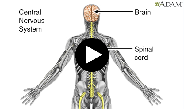
Have you ever wondered how your thoughts become actions? You think about raising your arm and suddenly your arm is raised. You think about jumping and before you know it your legs lift off the ground and come back.
In fact, there are many steps that happen in your body between thinking and action. The part of your brain that “thinks” about the action needs to communicate its intention to the part of the brain that controls movement. The signal to move is then sent from the brain to the spinal cord and out to the nerves in the relevant area of the body, which activates the necessary muscles to move.
Any obstacle along the way from thought to action can prevent the action from taking place. For example, brain or spinal cord injuries that affect signals from the brain mean that the signal to move cannot be communicated to the relevant area of the body. Damage or loss of a limb can prevent an action from taking place even if this signal is communicated. A new technology called the
brain-machine interface
or BMI (also called brain-computer interface or BCI) can enable thought to become action in people with these types of conditions or injuries.
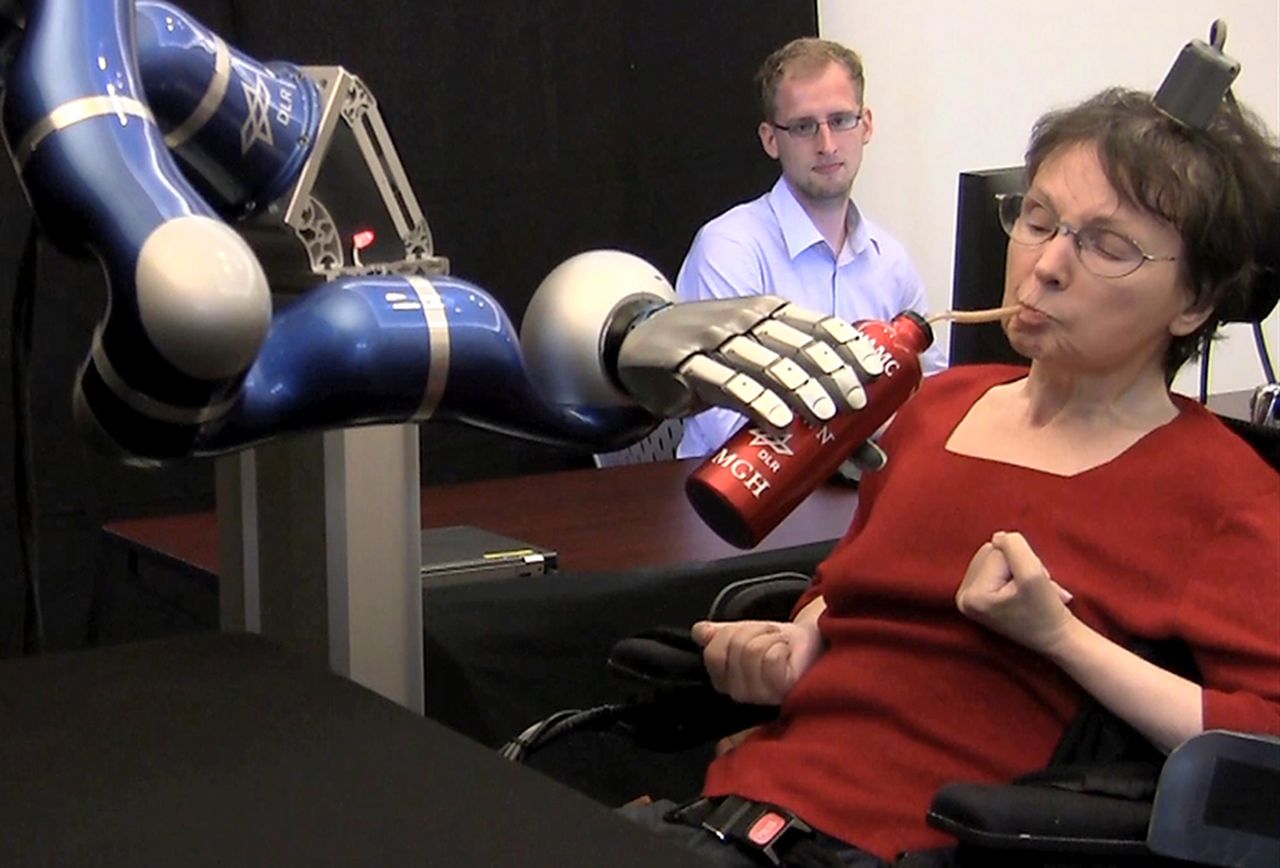
Figure 1. Woman using brain machine interface to bring a cup of coffee to her mouth via robotic arm
[Source: Image reprinted by permission from Macmillan Publishers Ltd: Nature, Copyright 2012. https://www.pnas.org/content/110/46/18343]
Dr. Kelly Clancy first became interested in BMIs while in graduate school in neuroscience. A friend had lost several limbs and the availability of technology such as
prosthetics
to support this friend to regain limb function were limited. Dr. Clancy turned to BMIs as a new frontier of neuroscience research, with the goal of supporting people like this friend in their journey to reconnect thought and action.
BMIs are more than a promising therapy for patients; they're also a tool that neuroscientists can use to better understand how the brain works. For people with spinal cord injuries, therapeutic options related to BMIs are limited. Available BMIs in humans require surgery to implant
electrodes
into the brain that can transfer electrical activity from the brain to a computer. The surgery can cause many complications, and the function of the electrodes may only last several months. The purpose of Dr. Clancy’s work with BMIs is to learn more about the brain processes that connect thought and action in order to develop new, safer technologies for patients.
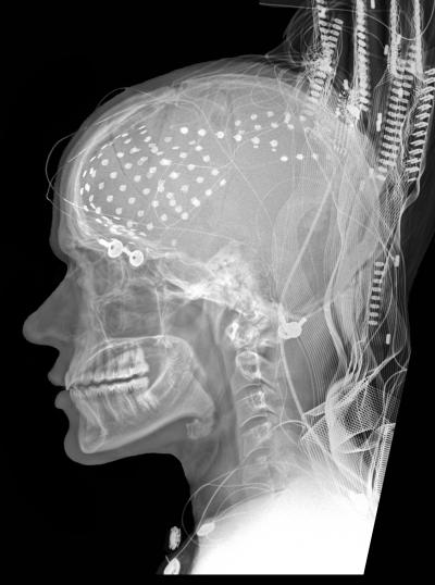
Figure 2. X-ray image of electrodes attached to the brain
[Source: Ned T. Sahin, Ph.D. http://www.nedsahin.com/research/media_images/]
Creating a Brain Machine Interface
Brain Machine Interfaces typically include three parts: a way to record brain activity (such as electrodes), a computer that interprets the electrical activity, and a task that needs to be completed. One example of a task for patient use is to enable a patient to type on a computer with their mind. In other words, they are able to type on the keyboard just by thinking about it. In animal experiments, BMIs usually involve a visual task such as moving a cursor on a computer screen.
Although electrodes are the most common method used to record brain activity, neuroscientists are experimenting with other techniques that are less invasive. Dr. Clancy used a technique called
widefield fluorescent microscopy.
Fluorescence is a technique used in neuroscience to visualize electrical activity in
neurons
(nerve cells). It is a property of proteins found in some marine animals, such as jellyfish, that glow in the dark.
These fluorescent proteins can be modified and inserted into the genetic material of other animals, like mice, so that certain cells (like neurons) will fluoresce. In particular, these proteins were engineered to become brighter when neurons are active: increased fluorescence in neurons can indicate electrical activity because the fluorescent proteins glow more brightly when exposed to calcium. Neurons have very low levels of calcium except when they are activated. As a result, the brighter the neurons fluoresce, the more electrical activity is happening.
For this experiment, Dr. Clancy used a mouse model that expressed the GCamp6s fluorescent jellyfish protein in a specific type of neuron called
pyramidal neurons.
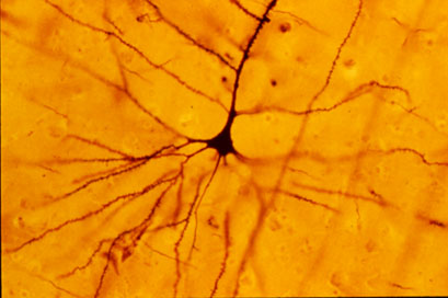
Figure 3. Pyramidal neuron
[Source: Bob Jacobs, Laboratory of Quantitative Neuromorphology, Department of Psychology, Colorado College. https://en.wikipedia.org/wiki/Pyramidal_cell#/media/File:GolgiStainedPyramidalCell.jpg]
Pyramidal neurons transform incoming information from a chemical form such as
neurotransmitters
into electrical activity. Widefield imaging of fluorescing pyramidal neurons allowed Dr. Clancy to observe brain activity in real time while the mice completed specific tasks.
To conduct these experiments, mice were trained on a visual task that required them to move a visual computer cursor from the center of the screen to a goal location at the edge of the screen using only their brain activity. The mice were given 30 seconds to complete the task. If they succeeded, they were given a reward of sugar water or soy milk. Whether they succeeded or failed, the task would reset after several seconds for the mouse to try again.
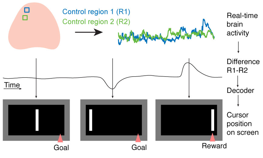
Figure 4. The mice were trained on a visual task that required them to move the cursor to the edge of the screen by activating control region 1 (R1) and deactivating control region 2 (R2)
[Source: Image courtesy of Dr. Kelly Clancy]
When mice are exposed to new tasks, they may randomly engage many different parts of the brain all at once as they try to figure out how to complete the task and gain their reward. The researchers did not want the task to be completed randomly as a result of a blast of brain activity. Rather, they wanted the mice to intentionally activate or deactivate designated brain areas.
For this reason, the researchers created a task that could be completed only by exerting control over specific areas of the brain. Activity in one region needed to be increased while activity in the other region was lowered. When this happened, the cursor would move to the goal location, and the mouse would receive a reward.
Throughout the course of the experiment, Dr. Clancy observed the brain activity of the mice under the microscope. She noticed the differences in brain activity as the mice became more familiar and eventually expert at the visual task.
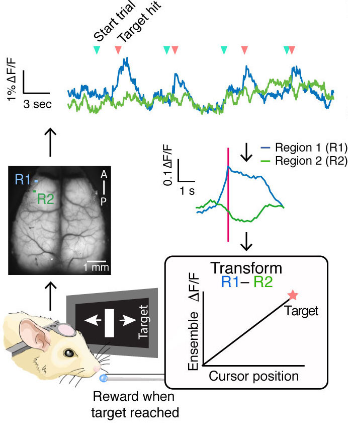
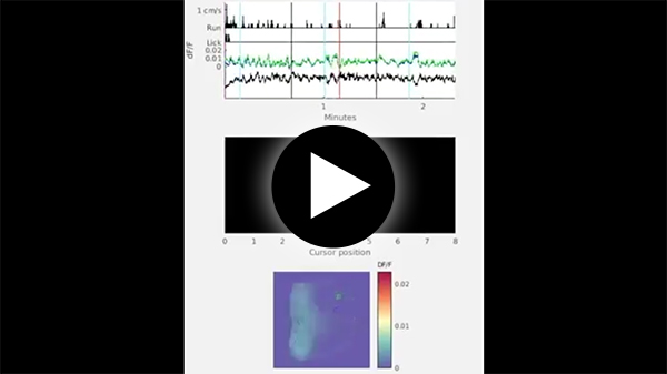
Figure 5. Experimental Brain Machine Interface developed by Dr. Kelly Clancy where mice are trained to complete a visual task by selectively activating and deactivating specific regions of the brain
[Source: Image courtesy of Dr. Kelly Clancy]
Dr. Clancy also observed the mice when they were watching the screen but not performing the task to see if there were differences in brain activity between passively watching the control task and actively doing it.
Deepening Our Understanding of the Brain
At the beginning of the experiment (before the mice learned the task), the mice had about a 25% chance of randomly using their brains to complete the task and receive a reward. Once the mice became experts, they were able to complete the task successfully about 70% of the time.
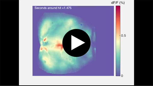
While observing the brain activity in the mice as they completed the task, Dr. Clancy noticed a difference in brain activity in the mice who were first exposed to the task and those who had become expert. When the mice were first exposed to the task, Dr. Clancy noticed the expected general increase in brain activity. In addition, she noticed heightened activity in the
primary visual cortex,
the part of the brain that receives visual information directly from the eye.
Figure 6. Location of primary visual cortex
[Source: https://en.wikipedia.org/wiki/Visual_cortex#/media/File:Visualcortex.gif]
Once the mice became better at completing the task, Dr. Clancy noticed increased activation of specific parts of the visual cortex that are related to processing the information received by the primary visual cortex and also involved in planning motor actions. This area of the mouse brain is related to the brain region in humans known as the
parietal cortex.
The parietal cortex is the outermost layer of the
parietal lobe,
one of the four main regions of the human brain.
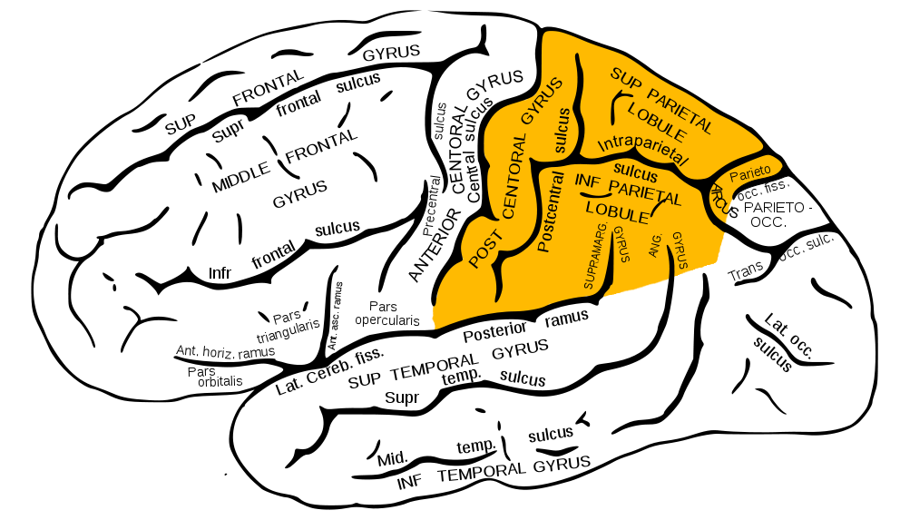
Figure 7. Parietal lobe highlighted in orange
[Source: https://en.wikipedia.org/wiki/Parietal_lobe#/media/File:Gray726_parietal_lobe.png]
In humans, the parietal cortex is thought to be related to a
sense of agency.
When you voluntarily lift your arm, you do so knowing that you are the one who intended to lift your arm and that you are doing it yourself. This is what “sense of agency” means. If someone else were to lift your arm, it might be the same movement, but the brain processes are different because you did not intend to lift your arm and you aren't the one doing the movement. In patients who have damage to their parietal cortex, even when they lift their arm themselves, it might feel like someone else is doing it. The finding of increased activity in the higher visual cortices in mice and its relation to the parietal cortex in humans is an important area of future research.
Another discovery the researchers made is that there are important differences in brain activity when the mice are actively completing the task compared to when they are passively watching the screen. When completing the visual task, the mice are much more focused on the position of the cursor than when passively watching. To better understand these differences, Dr. Clancy created a computer program that would guess the position of the cursor on the screen based only on the electrical activity in the mouse brain. Using this computer program, Dr. Clancy could better reconstruct the position of the computer cursor using only information from the brain activity when the animals were performing the task versus when they were passively watching.
In future research, Dr. Clancy plans to focus on understanding how mice are controlling activity in specific brain regions. In this study, the researchers showed that it is possible to observe changes in electrical activity as mice are completing the visual task. The next steps are to better understand how the mice are able to do this.
Dr. Kelly Clancy is a postdoctoral fellow in the laboratory of Dr. Tom Mrsic-Flogel at University College London. Dr. Clancy is a neuroscientist whose recent research focuses on improving scientific understanding of how the brain works with the goal of creating better therapies for neurological conditions. Dr. Clancy is also a writer whose work has appeared in many places including The New Yorker and Wired. When not in the laboratory, Dr. Clancy enjoys writing, drawing, and spending time outdoors.
Dr. Thomas Mrsic-Flogel is the director of the Sainsbury Wellcome Centre for Neural Circuits and Behaviours at University College London, and his lab focuses on understanding how the brain makes decisions by combining sensory information with previously learned knowledge.
For More Information:
- Clancy, K. and T. Mrsic-Flogel. 2021. “The sensory representation of causally controlled objects.” Neuron, 109(4): 677-689. https://doi.org/10.1016/j.neuron.2020.12.001.
To Learn More:
Brain Machine Interface
- Kelly Clancy's Website. https://www.kellybclancy.com/about
- Mrsic-Flogel Laboratory. https://www.sainsburywellcome.org/web/groups/mrsic-flogel-lab
Spinal Cord Injuries
- National Institute of Neurological Disorders and Stroke. https://www.ninds.nih.gov/disorders/all-disorders/spinal-cord-injury-information-page
- American Association of Neurological Surgeons. https://www.aans.org/en/Patients/Neurosurgical-Conditions-and-Treatments/Spinal-Cord-Injury
- Christopher and Dana Reeve Foundation. https://www.christopherreeve.org/living-with-paralysis/health/causes-of-paralysis/spinal-cord-injury
Written by Rebecca Kranz with Andrea Gwosdow, PhD at www.gwosdow.com
HOME | ABOUT | ARCHIVES | TEACHERS | LINKS | CONTACT
All content on this site is © Massachusetts
Society for Medical Research or others. Please read our copyright
statement — it is important. |