Dr. Ogle and her team created the first three-dimensional (3D) heart model, which allows for a fuller understanding of heart function and heart diseases.
This heart model was different from previous models because it used bio-ink containing stem cells and other proteins. The model was created using a 3D printer that printed the bio-ink layer by layer into a 3D model.
This 3D heart model was created to be the size of a mouse heart to be used in future experiments with mouse models. This will allow researchers to study progressive diseases of the heart.
In the early years of What A Year, over a decade ago, we highlighted a breakthrough in biomedical engineering: Dr. Kevin Kit Parker and his colleagues had engineered a two-dimensional strip of heart tissue. Prior to this development, it was difficult for researchers to test how different medications affected the heart and it was challenging to create models of diseased heart tissue to find better treatments for heart problems.
Over the past decade, researchers around the world have used two-dimensional models of heart tissue to create better and safer medications, develop better treatments for heart issues, and study diseases of the heart. However, the heart is a three-dimensional (3D) organ whose functions cannot always be studied using a two-dimensional model. For example, there are some disease states that progress due to the volume of blood in the heart and the amount of pressure the heart muscle can exert on the blood. The volume and pressure of a working heart can only be re-created with a 3D model. This is why biomedical engineers have been working to develop a 3D heart model to study the complex functions of the heart.
One common technique used to create three-dimensional models is
3D printing,
where a special printer prints out a 3D design layer by layer. Engineers working to create 3D-printed hearts have met with some success in creating a physical model of the heart. However, until now none of these heart models have been able to function the way the heart does. Recently, Dr. Brenda Ogle, graduate research students Molly Kupfer (who recently received her Ph.D.) and Wei-Han Lin, and their collaborators at the University of Minnesota have created the first-ever functional 3D heart model using a specialized biological ink (bio-ink) containing stem cells.
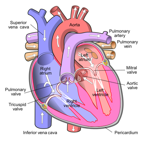
To understand how this 3D heart model works, let's review the function of the heart. Recall that the role of the heart is to circulate blood from the heart to the rest of the body and back to the heart. The blood that leaves the heart carries oxygen and other nutrients to the rest of the body, and once oxygen and nutrients have been used by organs and tissues, the blood comes back to the heart and travels to the lungs to be filled again with oxygen and nutrients. A healthy heart regulates its rhythm to maintain the right amount of blood in the heart and in the blood vessels.
There are a variety of cell types that work together to make the heart function. The inside of the heart and blood vessels are lined with a thin layer of cells called
endothelial cells
that assist in regulating blood vessel contraction and relaxation, blood clotting, and the immune response. About one-third of the heart has cells called
cardiomyocytes,
which are responsible for regulating the heart rhythm. Another cell type within the heart are
stem cells,
precursor cells to cardiomyocytes, which have not yet specialized into a certain type of cell. The process of specialization of stem cells is called
differentiation.
Epithelial cells, cardiomyocytes, stem cells, and all other cell types in the heart are held together by a complex web of proteins called the
extracellular matrix
or ECM.
In the past, researchers have developed 3D heart models with living heart tissue. A challenge these models have not been able to overcome is to make the heart cells work together as a pump, the way the human heart does. These models have used differentiated cardiomyocytes, which are known to be difficult to grow in the laboratory. Dr. Ogle and her team wondered whether they could achieve a functional heart chamber by using stem cells rather than adult cardiomyocytes. To test their hypothesis, they worked to develop a bio-ink that could be used to print 3D living tissue.
The base of the bio-ink was formed by the
hydrogels
called gelatin methacrylate (GelMA) and collagen methacrylate (ColMA). Hydrogels are water-swollen cross-linked polymers that create a mesh when chemically prompted. Dr. Ogle and her team performed several experiments to determine the optimum amount of GelMA in their bio-ink, which they found to be between 10% and 15%. To the GelMA base, the researchers added stem cells and ECM proteins that had previously been shown to support the differentiation of stem cells and
proliferation
of cardiomyocytes: fibronectin and laminin-111. The rest of the bio-ink was made of ColMA.
Dr. Ogle and her team created a variety of formulations of bio-ink, with the concentration of GelMA ranging between 10% and 15%, and varying amounts of fibronectin, laminin-111, and ColMA. Each trial bio-ink was placed into wells and monitored for growth of stem cells and cardiomyocytes. The results showed the optimal concentrations of each compound in the bio-ink, which was the formula that was then used for all future experiments.
For a 3D printer to work, it needs to have an image to print. The image that Dr. Ogle and her team used for printing was created by
magnetic resonance imaging
(MRI), a powerful imaging tool that visualizes soft tissue. The initial image was of a human heart, though it was scaled down to the size of a mouse heart for testing purposes, about 0.5 inches (1.3 cm) in diameter. The researchers also simplified the image to contain only one chamber with one blood vessel on either end. In contrast, an adult human heart has four chambers and multiple blood vessels that bring blood to and from the heart.
Once Dr. Ogle and her team had optimized the bio-ink and modified the image to print, it was time to try it out. The 3D printer printed one layer after another of live cells into the chambered structure Dr. Ogle had designed.
Figure 2. Researchers at the University of Minnesota have 3D printed a functioning centimeter-scale human chambered heart muscle pump in the lab. This video shows how the bio-ink containing cells and proteins is printed in a slurry bath. The entire process takes less than five minutes, but this video is at five-times the normal speed. [Credit: Kupfer, Lin, et al., University of Minnesota]
Next, Dr. Ogle applied chemical signals to promote differentiation of the stem cells. After two weeks, Dr. Ogle and her team found that over 90% of the bio-ink contained stem cells. Six weeks later, the effects of the chemical signals were measured. Dr. Ogle found that approximately 85% of stem cells had differentiated into cardiomyocytes, 10% into
smooth muscle
cells, and 4% into endothelial cells. The cells covered the entirety of the chamber and were several layers thick.
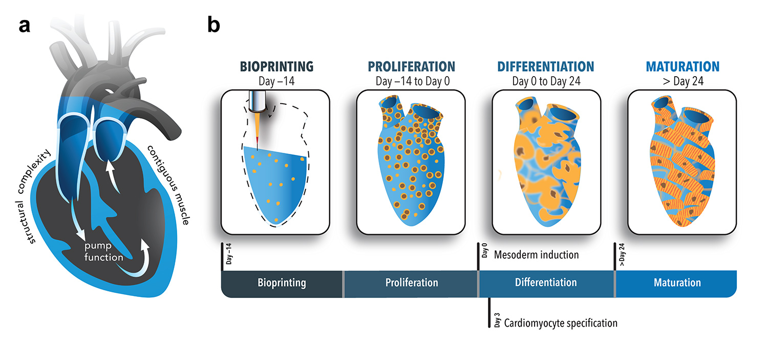
Figure 3. This figure illustrates the steps involved in generating the human heart pump. [Credit: Kupfer, Lin, et al., University of Minnesota]
In addition, the cardiomyocytes formed connections between each other that allowed them to transmit electrical signals and work together to pump fluid through the chamber.
Figure 4. The cardiac muscle cells of the human chambered muscle pump cover the majority of the structure as evidenced by visualization of the propagation of action potentials visualized here by bright white pixels that light up in sequence. [Credit: Kupfer, Lin, et al., University of Minnesota]
The researchers could observe the chambers beat on their own.
Figure 5. In a groundbreaking new study, researchers at the University of Minnesota have 3D printed a functioning centimeter-scale human chambered heart muscle pump in the lab. The pump can contract spontaneously and synchronously visible to the naked eye. [Credit: Kupfer, Lin, et al., University of Minnesota]
“It is unbelievable to watch these chambers beat on their own,” commented Dr. Ogle. “This is really a huge breakthrough in the field.” Indeed, the creation of a 3D beating heart-chamber is a huge step for researchers who want to study how the heart functions in healthy and diseased states and test different treatments. The model Dr. Ogle and her team developed is particularly unique because the bio-ink they created uses undifferentiated stem cells.
Figure 6. One of the unique qualities of the human chambered muscle pump is the unique geometry that allows for fluid perfusion of the engineered cardiac tissue. [Credit: Kupfer, Lin, et al., University of Minnesota]
There is still much more work to be done to optimize the heart chamber that Dr. Ogle and her team developed. For example, Dr. Ogle plans to improve the bio-ink to create a thicker muscle wall in the model and allow for the maturation of more cardiomyocytes. The 3D chamber was designed to be the size of a mouse heart to be used in future animal studies.
Dr. Brenda Ogle is Professor and Department Head of Biomedical Engineering, University of Minnesota College of Science and Engineering and Director of the Stem Cell Institute at the University of Minnesota. Her recent research focused on the differentiation of stem cells into cardiomyocytes, which has been a key factor in the development of a 3D printed heart chamber. When not in the laboratory, Dr. Ogle enjoys spending time with her family and being outdoors.
Dr. Molly Kupfer completed her Ph.D. in Biomedical Engineering in the laboratory of Dr. Brenda Ogle at the University of Minnesota, where she utilized human stem cells and 3D printing to generate living heart tissue. In her current job, she performs clinical evaluations of cardiac medical devices. Outside of work Molly enjoys drawing, doing the New York Times Crossword puzzles, and spending time with her cat.
Wei-Han Lin is currently a Ph.D. student in Biomedical Engineering at the University of Minnesota. Under the supervision of Dr. Brenda Ogle, his research merges engineering principles and 3D printing technology with stem cell biology to develop regenerative therapeutics and medical devices to treat heart disease. In his free time, Wei-Han enjoys cooking and watching ball games on TV.
For More Information:
Kupfer, M. et al. 2020. “In Situ Expansion, Differentiation, and Electromechanical Coupling of Human Cardiac Muscle in a 3D Bioprinted, Chambered Organoid.” Circulation Research, 127: 207-224. https://www.ahajournals.org/doi/10.1161/CIRCRESAHA.119.316155