| |
Highlights
Adolescence is a critical phase of life marked by improvements in cognitive, social, and emotional processing. Neuroscientists are beginning to see how these behavioral changes are associated with changes in the brain during this unique developmental period.
Neuroplasticity refers to the ability of the brain to change or reorganize based on experiences and the environment. These researchers introduced a new technique to measure neuroplasticity in the human brain and used this technique to quantify changes in neuroplasticity during development in children and adolescents ages 8 to 23 years. They found that neuroplasticity in the areas of the brain involved in visual and motor functions began to decline by age 8, whereas neuroplasticity in the areas of the brain involved in critical thinking, decision-making, emotional regulation, and social learning peaked around age 15.
Further studies showed that environmental factors can influence an adolescent’s window of neuroplasticity in brain regions supporting critical thinking and decision-making functions. Future research will conduct similar experiments in subcortical regions of the brain.
Adolescence is a critical phase of life typically marked by the physical changes of puberty and behavioral changes as young people seek more independence from their parents, engage with new environments, and become more sensitive to social relationships. This is a time of transition from childhood to adulthood, where adolescents begin to explore the world, contribute to their communities, and think more flexibly and abstractly. Previously thought to include just the teenage years – from ages 13 to 19 – this window has expanded based on developmental research to include young people ages 10 to 25 years old (and older!). New research by Valerie Sydnor, a doctoral student at the University of Pennsylvania School of Medicine in the laboratory of Dr. Theodore Satterthwaite and a large team of colleagues, shows that the changes of adolescence can also be seen in the brain.
The Developing Brain and Neuroplasticity
The size and functionality of our brains is what makes us human. When babies are born, their brains are about one-quarter the size of an adult brain. In the first year of life, the brain doubles in size. By age three years, the brain is an estimated 80% of adult size, and by age five years an estimated 90% of adult size. By early adolescence, the brain has reached its adult size. However, the parts of the brain involved in critical thinking, social cognition, and emotional regulation continue developing until a person is around 30 years old.
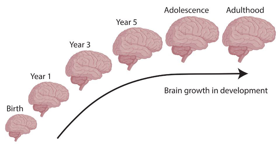
Figure 1. Brain growth from birth to adulthood.
[Source:Valerie Sydnor]
The building blocks of the brain are specialized nerve cells called neurons. Neurons transmit information to and from different regions of the brain and the body through electrical and chemical signals, a process referred to as neuronal communication or neuronal signal “firing”. Neurons that often transmit similar information get connected, or structurally wired, to each other to form
neural circuits.
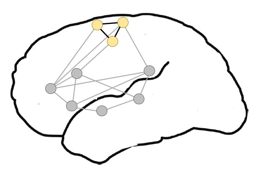
Figure 2. Neurons that often transmit similar information get connected to each other to form neural circuits.
[Source: https://en.wikipedia.org/wiki/Neural_circuit]
As a baby grows into a child, teenager, and adult, more neural circuits that encode experiences, memories, and habits are formed and then refined. The ability of neuronal circuits in the brain to re-wire, re-organize, or adapt their biological features is called
neuroplasticity.
In other words, neuroplasticity refers to the ability of the brain to change based on experiences and the environment.
To a certain extent, the human brain remains plastic our entire lives. This is how even older adults can regain function after a brain injury. However, it is well known that plasticity decreases with age. In general, this is why it is easier for children to learn new things—such as a second language—than adults. The youth brain is specifically biologically programmed to have enhanced development-related neuroplasticity.
Structures of the Brain
The brain can be divided into three main parts: the
brain stem,
the
cerebellum,
and the
cerebrum.
The brain stem connects the brain to the spinal cord and controls many automatic processes that are essential for life, such as breathing and heart rate. The cerebellum sits below the cerebrum and is responsible for posture and balance. The cerebrum, the largest part of the brain, regulates all other functions, including interpretation of visual, auditory, and other sensory information, language, reasoning, learning, decision making, and processing of social and emotional information.
The cerebrum is divided into four main regions, called lobes, that are associated with different functions:
- Frontal lobe: associated with decision-making, emotional control, personality, and speech production.
- Parietal lobe: associated with spatial understanding and the integration of different types of sensory information.
- Occipital lobe: associated with processing visual information including object and facial recognition and depth perception.
- Temporal lobe: associated with hearing, language, and memory.
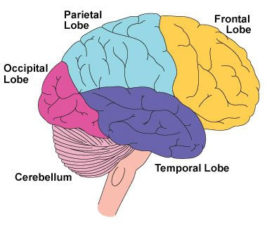
Figure 3. Four main regions of the cerebrum.
[Source: https://en.wikipedia.org/wiki/Frontal_lobe_disorder#/media/File:Frontal_lobe.png]
Information in all the lobes is processed by the outermost half-inch of the cerebrum, known as the
cerebral cortex.
The cerebral cortex is divided into three areas based on function: sensory, motor, and association.
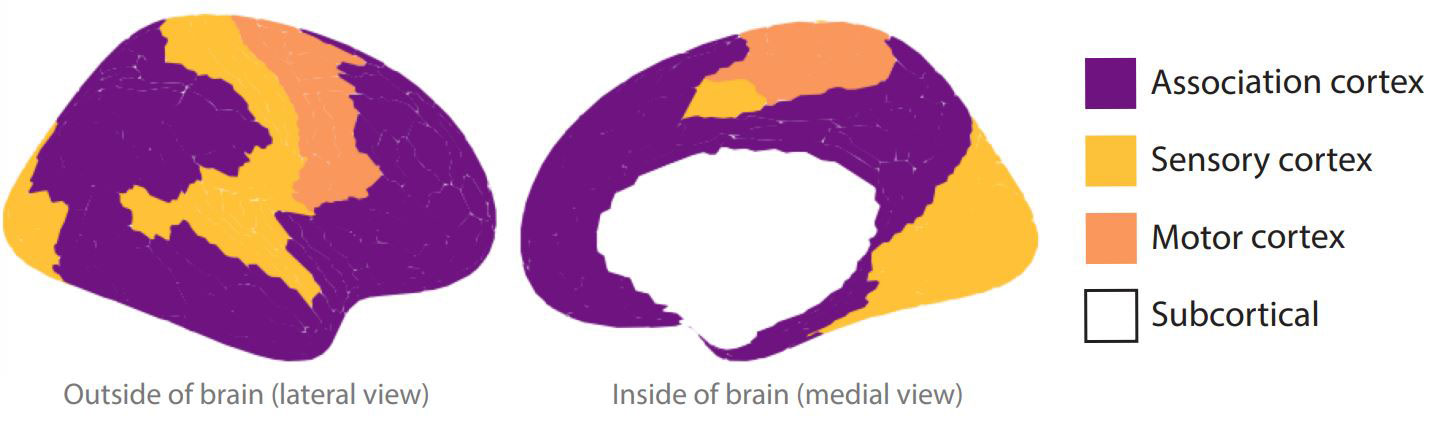
Figure 4. Sensory, motor, and association areas of the cerebral cortex. Subcortical structures (in white) are located deeper in the brain and are not part of the cerebral cortex.
[Source: Source: Valerie Sydnor]
Sensory function refers to how the cerebral cortex interprets information related to sight, hearing, touch, taste, and smell. Motor function refers to how the cerebral cortex controls and coordinates voluntary muscle movements. Sensory and motor functions are often grouped together in what is called the sensory-motor cortex. Association functions refer to higher-order processing that gives meaning and context to information. For example, the sensory-motor cortex processes visual information so that you see a clear picture, while the association cortex provides the interpretation of what is seen.
Developing a Measure of Neuroplasticity
Valerie joined the Lifespan Informatics and Neuroimaging Center at the University of Pennsylvania, which Dr. Satterthwaite leads, to better understand how the brain develops, how brain development is impacted by the environment, and how changes in brain development can increase one’s risk for mental health conditions such as
depression,
schizophrenia,
and
bipolar disorder.
Many mental health conditions appear during adolescence, and the longer someone goes without treatment, the more difficult it can become to treat. This speaks to the ways that our brains tend to become less plastic—less open to change—over time. “I joined Dr. Satterthwaite’s lab because I wanted to figure out how we could tailor psychiatric interventions to the times in brain development when they are most likely to be effective and most likely to have a lasting beneficial impact on the brain,” Valerie recalled. This would require a more nuanced understanding of brain development and plasticity.
Although neuroplasticity is a key concept that underpins our understanding of the brain, there was no quantitative measure of neuroplasticity in humans prior to Valerie’s research. Developing an objective and biologically informed readout of neuroplasticity became one of Valerie’s project goals.
Working together with Dr. Bart Larsen, a post-doctoral researcher in Dr. Satterthwaite’s laboratory, Valerie scanned the neuroscience literature for clues. Most of the literature described experiments that had been conducted in rodent models. These experiments identified specific cells and molecules that were involved in regulating neuroplasticity, but there was no way to measure these markers in humans. Instead, Valerie and Dr. Larsen were looking for a measure of brain activity that was linked to neuroplasticity and could also be seen using currently available human brain imaging techniques.
Insight from animal experiments showed that when an area of the cerebral cortex is more plastic, neurons in that area tend to show a different pattern of neuronal communication, or firing. Specifically, when an area of the cerebral cortex is more plastic, neurons fire more spontaneously and more in synchrony than in an area that is less plastic. The researchers hypothesized the relative strength and synchrony of spontaneous neural activity in a given area of the brain would indicate whether that area was more or less plastic. They therefore determined how to study this measure of activity-indexed neuroplasticity in the human brain using a safe form of brain imaging called functional magnetic resonance imaging (fMRI). Once they developed this technique, they were able to use this quantitative measurement of neuroplasticity in subsequent research.
Analyzing the Philadelphia Neurodevelopmental Cohort
Valerie, Dr. Larsen, and Dr. Satterthwaite applied this new metric of neuroplasticity to 1,033 publicly available brain scans of adolescents 8 to 23 years obtained from the Philadelphia Neurodevelopmental Cohort (PNC). The PNC is an ongoing collaboration between the Brain Behavior Laboratory at the University of Pennsylvania and the Center for Applied Genomics at Children’s Hospital of Philadelphia. The dataset includes over 9,500 young people ages 8 to 23 years who received care at the Children’s Hospital of Philadelphia and agreed to participate in this research.
As part of their participation in the PNC, 1,601 young people volunteered to undergo
functional magnetic resonance imaging (fMRI)
while simply resting and awake in an MRI scanner. Resting fMRI is a non-invasive imaging study that allows researchers to look at the strength and synchrony of spontaneous activity in different parts of the brain. During an fMRI, patients are placed on a table and lie still inside a magnetic tube, just like a standard MRI.
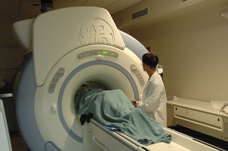
Figure 5. A patient entering an MRI machine.
[Source: https://en.m.wikipedia.org/wiki/File:MRI_machine_with_patient_(23423505123).jpg]
It takes a large team of specialists to develop the software needed to analyze brain scans from an MRI. Valerie collaborated with Dr. Azeez Adebimpe, Sydney Covitz, and Dr. Matthew Cieslak from Dr. Satterthwaite’s bioinformatics team to develop the code needed to analyze the brain scans and determine neuroplasticity measurements for different regions of the brain. They used scans from a total of 1,033 participants after excluding scans with low quality and scans from participants who had underlying medical conditions.
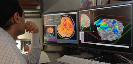
Figure 6. Image of a researcher analyzing fMRI scans.
[Source: https://en.wikipedia.org/wiki/Functional_magnetic_resonance_imaging]
The researchers divided the brain into 336 cortical regions and coded each region according to where it lies on the sensory-motor to association axis of the cerebral cortex. This spatial axis captures the extent to which a cortical region is necessary for perception or movement related functions versus advanced mental functions such as cognitive control, reasoning, planning, social understanding, and emotional regulation.
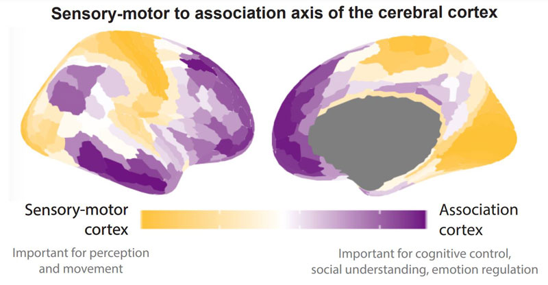
Figure 7. The researchers divided the brain into 336 cortical regions and coded each region according to where it lies on the sensory-motor to association axis of the cerebral cortex.
[Source: Valerie Sydnor]
For each brain scan, the researchers quantified the fMRI index of plasticity within each of the 336 cortical regions. Then they looked across scans to study how this measure changed with age in each region. They were able to characterize a trajectory of neuroplasticity in all cortical regions from ages 8 to 23 years. The results showed that neuroplasticity in the sensory-motor cortex was already declining by age 8 years and continued to decline until early-to-middle adolescence. However, neuroplasticity in the association cortex continued to increase from age 8 to its peak at age 15 years before then declining into early adulthood.
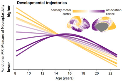
Figure 8. Developmental trajectories of neuroplasticity across development. Neuroplasticity in the sensory-motor cortex (yellow) was already declining by age 8 and continued to decline until early-to-middle adolescence. Neuroplasticity in the association cortex (purple) increased from age 8 to its peak at age 15 years before declining into early adulthood.
[Source: Valerie Sydnor]
These results support previous evidence that adolescent brains continue to develop well into the teenage years, and even suggest an enhancement in plasticity in areas important for cognitive, social, and emotional processing.
Brain Development and Your Environment
Next, the researchers wanted to know how children’s neurodevelopment might be impacted by their environment. Previous research has shown that differences in economic resources in the community where a child lives can impact brain development. Specifically, prior research seemed to indicate that children from neighborhoods with fewer socioeconomic resources may have a faster rate of brain development compared with children from communities with more resources. Valerie and her team sought to apply their new measure of neuroplasticity to see if they could identify brain changes based on the resource level and the socioeconomic circumstances of a child’s community.
To do this, the researchers used data from the PNC to create a neighborhood level factor score that would assign a numeric value to a child’s environment. This score was based on neighborhood census data obtained from participants’ zip codes, including:
- Neighborhood high school completion rate
- Neighborhood employment rate
- Neighborhood poverty rate
The researchers associated each brain scan with the corresponding neighborhood level factor score and analyzed the results to explore any differences. The results showed differences consistent with reduced plasticity in the association areas of the brain across all ages in young people with a lower neighborhood level factor score, a general approximation of a neighborhood with fewer resources. These findings provide further evidence for the hypothesis that neighborhood environment impacts neuroplasticity. The relationship between the neighborhood level factor scores and the fMRI measure of plasticity was strongest in the higher-level association areas of the brain.
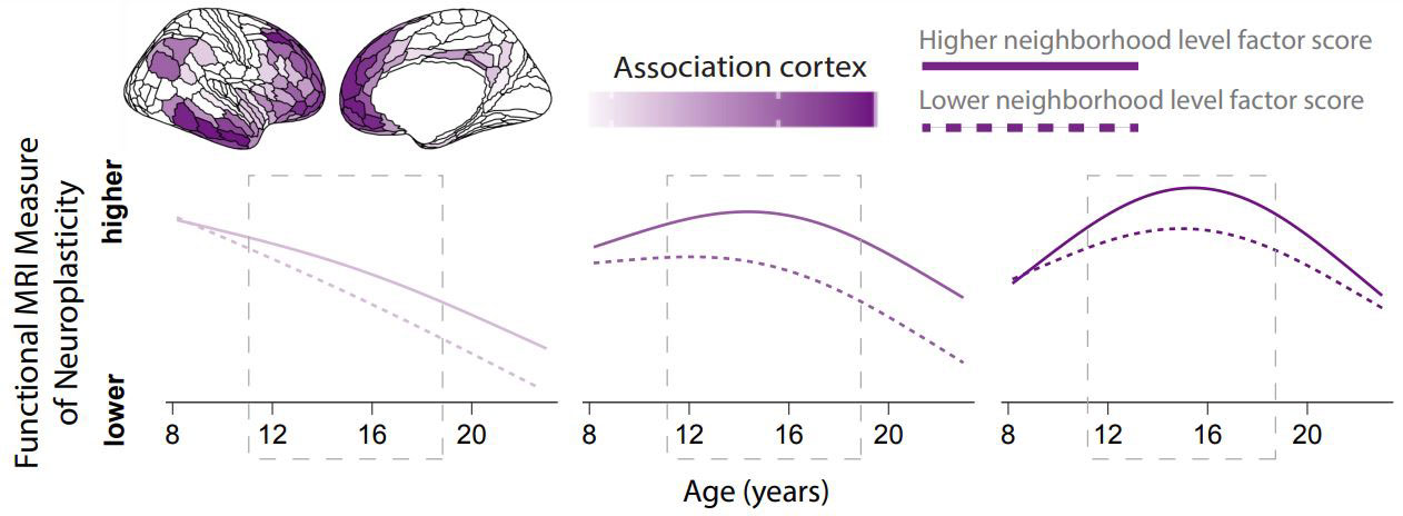
Figure 9. Comparison of neuroplasticity measure in the association cortex of young people with a higher neighborhood level factor score (solid line) and those with a lower neighborhood level factor score (dashed line). The results showed differences consistent with reduced plasticity in the association areas of the brain across all ages in young people with a lower neighborhood level factor score, a general approximation of a neighborhood with fewer resources.
[Source: Valerie Sydnor]
The most notable differences in neuroplasticity based on environmental factors occurred during adolescence.
Future Research
Many of you who are reading this article might be going through adolescence right now. “I want you to know that some of the changes you are experiencing are changes that we can see by studying the uniqueness of the adolescent brain,” explained Valerie. “Adolescence is a time when association regions of your brain remain particularly plastic—or malleable—such that the environments and contexts you explore can have a greater impact on them than at other times in your life.” In other words, because, as this research emphasizes, different parts of the brain mature at different times, the same experiences you have in adolescence—when your brain is more malleable—versus adulthood—when your brain is less malleable—can have different impacts. “While this may seem daunting at first”, Valerie noted, “I hope you can see adolescence as a developmental period characterized by opportunity. Adolescent brain plasticity allows this stage of life to be defined by learning, socioemotional transformation, and adaptive change.”
This research focused on the brain regions of the cerebral cortex, the outer layer of the brain responsible for a large diversity of human functions. In her next research projects, Valerie will use the same validated measure of neuroplasticity described here to further understand individual differences in cerebral cortex plasticity during development, as well as to understand interactions between plasticity in the cortex and the development of
subcortical structures.
Subcortical structures tend to be evolutionarily older parts of the brain that are structurally connected to specific areas of the cerebral cortex.
In future research, Valerie also hopes to explore the biological mechanisms driving increased plasticity during adolescence. Ultimately, Valerie hopes these research findings will be used to inform the timing and the type of targeted interventions for psychiatric illnesses in adolescents. For example, testing specific interventions at different times during adolescence based on our understanding of neuroplasticity to see if there is a time window at which an intervention is most successful.
Valerie Sydnor is a doctoral researcher in the laboratory of Dr. Theodore Satterthwaite at the University of Pennsylvania School of Medicine. Her research focuses on the neurodevelopmental basis of psychiatric illness risk. When not in the laboratory, Valerie enjoys running marathons, being outdoors, listening to musicals, and reading and watching fantasy.
Dr. Theodore Satterthwaite is the McLure Associate Professor of Psychiatry at University of Pennsylvania School of Medicine and Director of the Penn Lifespan Informatics and Neuroimaging Center, a research team comprised of neuroscientists, doctors, psychologists, statisticians, and computer scientists. His research focuses on using advanced neuroimaging and computational techniques to better understand neurodevelopment and the origins of psychiatric illness in youth. His lab also creates new methods and software to analyze MRI images and provides these as tools to the scientific community. When not in the laboratory, Dr. Satterthwaite enjoys biking, hiking, eating hot sauce, and spending time with his three children, who are rapidly approaching adolescence.
For More Information:
- Sydnor, V. et al. 2023. “Intrinsic activity development unfolds along a sensorimotor–association cortical axis in youth.” Nature Neuroscience, 26: 638-49. https://pubmed.ncbi.nlm.nih.gov/36973514/
- Sydnor, V. et al. 2021. “Neurodevelopment of the association cortices: Patterns, mechanisms, and implications for psychopathology.” Neuron, 109: 2820-46. https://pubmed.ncbi.nlm.nih.gov/34270921/
- Moore, T. et al. 2016. “Characterizing social environment’s association with neurocognition using census and crime data linked to the Philadelphia Neurodevelopmental Cohort. Psychological Medicine, 46(3): 599-610. https://www.ncbi.nlm.nih.gov/pmc/articles/PMC7263021/
To Learn More:
- Dr. Satterthwaite’s laboratory. https://www.pennlinc.io/research
Adolescence and Development
- UCLA Center for the Developing Adolescent. https://developingadolescent.semel.ucla.edu/core-science-of-adolescence/key-concepts
- Cleveland Clinic. https://my.clevelandclinic.org/health/articles/7060-adolescent-development
- U.S. Department of Health and Human Services, Office of Population Affairs. https://opa.hhs.gov/adolescent-health/adolescent-development-explained
- American Academy of Pediatrics. https://www.healthychildren.org/English/ages-stages/teen/Pages/Stages-of-Adolescence.aspx
- Raising Teens Project. https://hr.mit.edu/static/worklife/raising-teens/ten-tasks.html
Psychiatric Illnesses
- National Institute of Mental Health. https://www.nimh.nih.gov/health/statistics/mental-illness#part_2632
- National Alliance on Mental Illness. https://www.nami.org/About-Mental-Illness/Mental-Health-Conditions
- Centers for Disease Control and Prevention, Division of Adolescent and School Health. https://www.cdc.gov/healthyyouth/mental-health/index.htm
- U.S. Department of Health and Human Services, Office of Population Affairs. https://opa.hhs.gov/adolescent-health/mental-health-adolescents
Written by Rebecca Kranz with Andrea Gwosdow, PhD at www.gwosdow.com
HOME | ABOUT | ARCHIVES | TEACHERS | LINKS | CONTACT
All content on this site is © Massachusetts
Society for Medical Research or others. Please read our copyright
statement — it is important. |
|
|

Valerie Sydnor

Dr. Ted Satterthwaite

Valerie Sydnor and Dr. Azeez Adebimpe

Valerie Sydnor and Dr. Bart Larsen
Sign Up for our Monthly Announcement!
...or  subscribe to all of our stories! subscribe to all of our stories!

What A Year! is a project of the Massachusetts
Society for Medical Research.
|
|
