Itching, like pain, is a survival mechanism that draws the brain’s attention. Cholestatic itching is associated with liver disease and can be debilitating and significantly affect a patient’s quality of life.
These researchers identified a signaling pathway for cholestatic itch that reveals new targets for treatment. This pathway demonstrates for the first time that skin cells can act as “pre-neurons” in the context of human disease.
Next steps for this research include further elaborating critical details of this signaling pathway and designing and implementing new treatments for cholestatic itch.
Bitten by a mosquito? Don’t scratch! Mosquito bites can be very itchy, but did you know that scratching makes them worse? That’s because the sensation of itching is caused by your own body as it reacts to saliva from the mosquito and activates the immune system to neutralize the foreign agent.
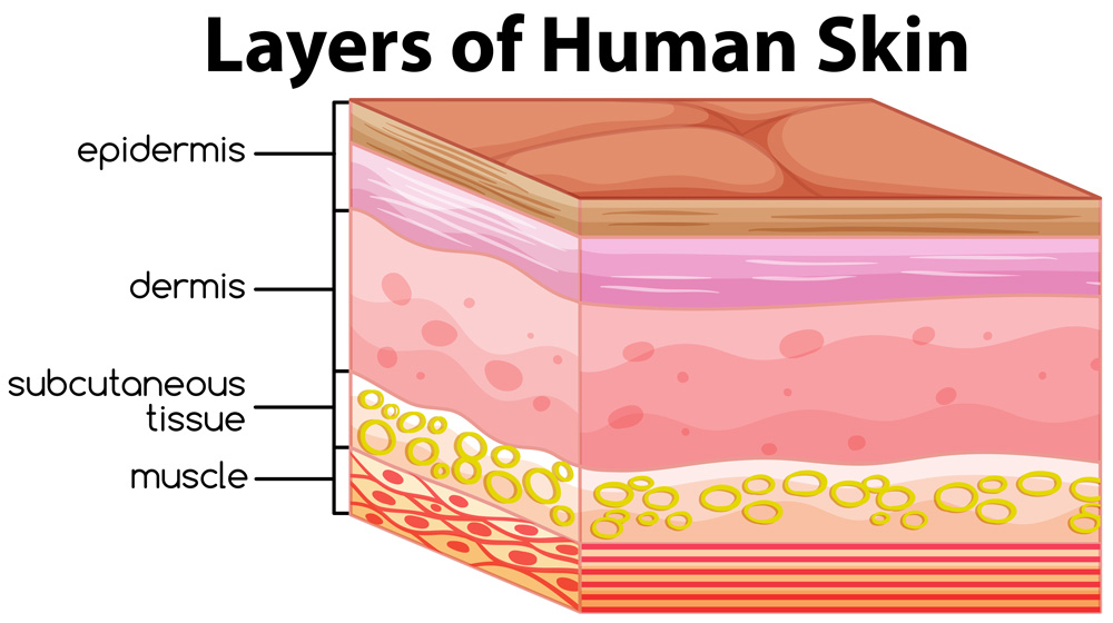
Itchiness, like pain, is an important survival mechanism that alerts our brains to something off or abnormal in the immediate environment surrounding the skin. Non-excessive (moderate) scratching accelerates healing of wounds. This is one of the many important functions of the skin, the largest organ in the human body. Other functions include acting as a watertight barrier to protect our internal organs from the outside environment, regulating body temperature, and allowing for our sense of touch, temperature, and external pain.
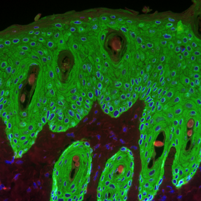
The primary role of keratinocytes is to form the outer layers of an organism that functions as a barrier between the inside and outside of our bodies. Keratinocytes keep out potentially dangerous environmental elements like water, dust, and ultraviolet (UV) rays from the sun. They also function as part of the
immune system
to protect against potential threats like bacteria, viruses, and parasites. When keratinocytes come in contact with such a threat, they can trigger the immune system to send specialized cells and extra fluid to neutralize the foreign element. This is what happens when you get bitten by a mosquito or have an allergic reaction.
Recently, scientists have shown the function of keratinocytes is not limited to the immune system and
inflammation.
In their ground-breaking research, Dr. Wolfgang Liedtke and colleagues from around the world have shown that keratinocytes also function as “pre-neurons,” functioning up-stream of nerve cells in the skin to send itch signals originating in the skin to the brain.
Pain and Itch
Dr. Wolfgang Liedtke, Chair of
Neurology
at Regeneron Pharmaceuticals, Adjunct Professor of Neurology at Duke University, and Adjunct Professor of Dentistry at New York University, is a clinician and researcher who has focused his career on helping patients experiencing chronic pain and other neurological problems. Two decades ago, his research identified a novel calcium-permeable
ion channel
which he suggested to be involved in pain.
This ion channel is now referred to as transient receptor potential vanilloid-type 4 or TRPV4. Since then, researchers have been studying how TRPV4 causes pain, with the goal of developing new therapeutic approaches.
In a study conducted several years ago, Dr. Liedtke and his colleague, Dr. Carlene Moore in the Liedtke Laboratory at Duke University, showed that TRPV4 in keratinocytes was responsible for pain caused by sunburn.
In this study, the researchers hypothesized a mechanism for how TRPV4 is involved in the sensation of itch. “Anyone who has experienced a sunburn knows it can be both painful and extremely itchy, with itch typically before and after pain,” commented Dr. Liedtke. “Our data have suggested that the same ion channel, TRPV4, in skin keratinocytes regulates these sensations when skin is burned by UVB rays.”
As a result of this finding, Dr. Liedtke and his team expanded their research on TRPV4 to include itching. As a first step, they focused on itching caused by
histamine.
This is the common type of itching that comes from a mosquito bite or allergic reaction. Dr. Liedtke was able to use
mouse models
in this study because mice also experience itching, which can be measured. When mice are injected with histamine into their skin, they show a reproducible scratching response that indicates itch. The researchers quantified the itching by recording the mice by video camera after a histamine injection and they counted how many times the mouse scratched an itch over a given time period.
Figure 3. Video of mice scratching. In this video, the mouse on the left has genetic changes that make it “immune” to the effects of chloroquine, which can cause severe itching in mice. The mouse on the right shows a normal mouse itch response. [Source: https://www.youtube.com/watch?v=oJAfR4e9k-Y&t=14s]
To determine the role of TRPV4 in itching caused by histamine, the researchers first exposed mice to histamine to provoke itching and then used a compound known to temporarily block the function of TRPV4. After itching due to exposure to histamine, mice that received the TRPV4 blocker temporarily stopped itching. The researchers found they could use the TRPV4 blocker to turn the histamine-provoked itching on and off, which showed that TRPV4 mediated the itch response in these mice.
To confirm these results, Dr. Liedtke and his lab member Dr. Yong Chen conducted a follow-up experiment using a
knock-out mouse
model that lacked the TRPV4 gene only in the skin. “This is a very specific animal model,” explained Dr. Liedtke. “TRPV4 is found in cells all over the body. We were interested only in TRPV4 in the skin.” In this experiment, two groups of mice, the control mice and the knock-out mice lacking TRPV4 in skin cells, were exposed to histamine to provoke itching. The control mice were observed to experience severe itching, as the researchers expected. In the knock-out mice, however, histamine did not provoke the itching response. From these findings, Dr. Liedtke concluded that TRPV4 in skin keratinocytes was responsible for histamine-related itching in mice.
“This was an exciting finding,” recalled Dr. Liedtke, “but it does not have truly relevant clinical applications, nor does it explain how the skin keratinocyte talks to sensory neurons in the skin.” Available antihistamines are quite safe and effective, and you can buy them over-the-counter in cream or pill form, depending on the severity and the location of the itch. While annoying, histamine-related itch is usually temporary, can be controlled, and does not chronically affect a patient’s quality of life. For these reasons, Dr. Liedtke decided to study a more serious form of itching called
cholestatic itching.
Cholestatic Itching
Cholestatic itching is associated with problems of the liver. The specific mechanisms that cause cholestatic itching are unknown, and there are no treatment options available. For patients with unbearable cholestatic itch, the only option for itch relief is an
organ transplant,
an extreme treatment that is only done as a last resort. Any treatment that could control itching for these patients would be enormously beneficial.
Previous research by Dr. Liedtke’s colleague Dr. Andreas Kremer, now heading Clinical Hepatology at University Hospital of Zurich in Switzerland, showed that itching in patients with cholestatic itch was associated with elevated levels of lysophosphatidic acid (LPA) in their blood. The level of LPA was remarkably correlated with itch intensity. Because of this finding, Dr. Liedtke and his team exposed mice to LPA in the laboratory to see whether it would provoke an itching response. They found that exposure to LPA evoked moderate itching in mice.
Next, the researchers decided to try a molecule that is a precursor to LPA called lysophosphatidylcholine (LPC). Previous research has demonstrated that LPC is present systemically and in skin lesions in conditions with itching such as
psoriasis
and
atopic dermatitis,
also known as
eczema.
When Dr. Chen, now with his own laboratory, and Dr. Liedtke injected mice with LPC into their skin, they found that it evoked a strong itching response. This itching response was much weaker in mice who lacked the TRPV4 gene in their skin cells, meaning that the ability of LPC to cause itch was critically hinging on the TRPV4 ion channel in the skin.
To better understand the potential role of LPC in cholestatic itch, Dr. Liedtke and his colleagues at Duke University partnered with Dr. Kremer and colleagues in Germany and Dr. Piotr Milkiewicz and colleagues in Warsaw, Poland. Drs. Kremer, Milkiewicz and colleagues conducted a study of LPC in patients with
primary biliary cholangitis,
a chronic liver disease that often causes cholestatic itch.
Participants with primary biliary cholangitis who agreed to give blood samples for this study were asked to quantify their level of itching on a scale from 0 to 10, with 0-3 meaning no/mild itch, 3-6 meaning moderate itch, and 6-10 meaning severe/worst imaginable itch.
The researchers found that patients with primary biliary cholangitis had elevated levels of LPC in their blood. Furthermore, the intensity of the itch corresponded statistically with the level of LPC: the higher the level of LPC, the greater the intensity of itch reported by patients.
In another large collection of primary biliary cholangitis patients from Duke University, Dr Liedtke and his
hepatology
colleagues, Drs. Anna Mae Diehl and Manal Abdelmalek, demonstrated that LPC was present in all patients regardless of whether they had underlying liver problems. From these findings, the researchers concluded the presence of some level of LPC in the blood did not necessarily correlate with itch. This means that there are other factors in addition to LPC that facilitate LPC-evoked itching in primary biliary cholangitis. Some of these factors, like
bilirubin
and
bile acid,
have been suggested in previous research, but these liver-gall bladder metabolites do not correlate with itch intensity.
Keratinocytes as “pre-neurons”
As the human clinical samples suggest, LPC is a powerful and clinically relevant itch-inducing lipid molecule that appears central to how cholestatic itch functions. To close the gap between experiments in mice, human cells, and the suggestive data from patients' blood, Dr Liedtke collaborated with Dr. Mei-Chuan Ko of Wake Forest Baptist Medical Center, a leading primate researcher. They found that all itch-inducing molecules (called
pruritogens)
from his study also evoked equally robust itching in rhesus macaques.
With further confidence about the validity of their data, the researchers then attempted to deconstruct the mechanism for how LPC activates the TRPV4 ion channel in skin keratinocytes, and how the activation of TRPV4 stimulated itch-sensing nerve cells in the skin. When going down this path experimentally, they identified a molecule known as microRNA-146a (miR-146a) released by skin keratinocytes. The researchers showed that miR-146a was a potent itch-inducing molecule when injected into the skin of mice and monkeys.
miR-146a is a
microRNA.
microRNA or miRNA are a more recent discovery in the field of genetics. They are short sequences of genetic material that function to turn on and off gene expression. What Dr. Liedtke discovered, however, is that miR-146a exerted its itch-inducing effect in a radically different manner, namely it functions as a signaling molecule to directly and immediately cause itch, similar to histamine or serotonin, not taking a detour via regulation of genes.
Importantly, miR-146a had previously been shown to be related to atopic dermatitis (eczema) and skin disease, and it was thought to be present as these skin conditions were healing. “This makes a lot of sense,” explained Dr. Liedtke, “because physiologic itch contributes to skin healing.”
Contributions to the Field
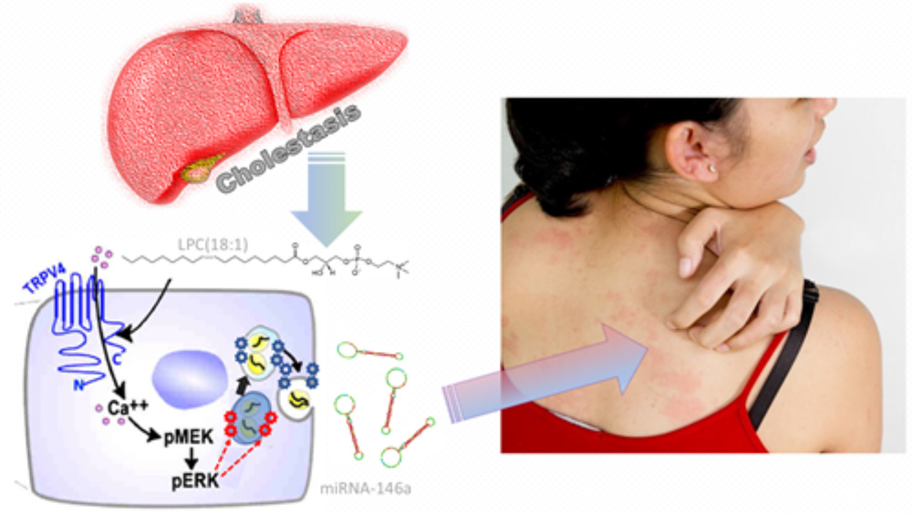
Through these experiments, Dr. Liedtke and colleagues described a new mechanism behind cholestatic itch, described below and shown in Figure 4:
Liver disease causes elevated levels of lysophosphatidylcholine or LPC
LPC directly activates calcium-permeable TRPV4 ion channels in skin keratinocytes
TRPV4 stimulates keratinocytes to release miR-146a
miR-146a causes the sensation of itch by directly activating sensory itch neurons
Figure 4. The mechanism behind cholestatic itch in the epidermis. Liver disease (cholestasis) causes elevated levels of LPC, which activate TRPV4 ion channels in skin keratinocytes. TRPV4 stimulates keratinocytes to release mi-146a, which causes the sensation of itch. [Source: Dr. Liedtke]
“As a neurologist, this is an incredibly surprising and thrilling result,” commented Dr. Liedtke. This is because neurologists study neurons, the specialized cells of the nervous system that are thought to be the dominant cell that can communicate directly from one cell to another. However, in the pathway described above, keratinocytes, not neurons, are the primary itch generating cells. For this reason, Dr. Liedtke refers to TRPV4-expressing keratinocytes as acting as “pre-neurons” in causing cholestatic itch.
This work is important because it suggests a number of new target molecules for treatment of unbearable cholestatic itch: TRPV4 in skin keratinocytes, intracellular keratinocyte signaling machinery that is key for release of miR-146a, LPC and miR-146a, and TRPV1 ion channels on itch neurons in the skin. Such new targets can help control itching in patients with liver disease to improve their quality of life and avoid an organ transplant. Beyond cholestatic itch, itching in patients with chronic kidney disease and itch-dominant psoriasis might also rely on LPC as a key mechanism of itch.
Future Research
There are many avenues for future research based on these findings. First, there is much more to learn about keratinocytes and how they function as “pre-neurons.” Second, the mechanism of cholestatic itch described above still needs to be extended with further laboratory experiments focused on communication between keratinocytes and sensory neurons in the skin. Third, the development of treatments for cholestatic itch based on these findings will require further translational-medical research including studies in humans.
Dr. Liedtke, formerly Professor of Neurology, Anesthesiology, and Neurobiology at Duke University (Durham, NC), now serves as Chair of Neurology, Psychiatry, Pain Medicine and Ophthalmology at Regeneron Pharmaceuticals (Tarrytown, NY). He continues on a dedicated mission to improve the lives of patients with chronic pain and itching toward safe and effective treatments. One focus of his research career has been to study TRPV4 and other signaling pathways involved in chronic pain, inflammation, and tissue injury. Outside of his work. Dr. Liedtke enjoys music, spending time with his family, traveling, bicycling, hiking, exercising, and reading.
Dr. Liedtke’s work is very collaborative. Dr. Liedtke worked with many investigators for this research. Dr. Yong Chen is Assistant Professor of Neurology at Duke University who continues the study of the neurobiology of pain and itch in his laboratory with a focus on TRP ion channels. Dr. Ru-Rong Ji, Professor Anesthesiology, Neurobiology, and Cell Biology at Duke University, is a global authority on pain research. Dr. Andrea Nackley, Associate Professor of Anesthesiology at Duke University, is a pain researcher with particular focus on sex differences in pain research. Dr. Jennifer Zhang, Associate Professor of Dermatology at Duke University, is a skin researcher with deep expertise in keratinocyte biology. Dr. Anna Mae Diehl is Professor of Medicine at Duke University whose research focuses on liver injury and repair. Dr. Manal Abdelmalek, Professor of Medicine at Duke University, specializes in risk factors and treatment for liver disease. Dr. Mei-Chuan Ko, Professor of Physiology and Pharmacology at Wake Forest University, is a leading primate sensory researcher. Dr .Tony Reeves, Assistant Professor of Molecular Medicine at Wake Forest University, is a computational biologist. Dr. Andreas Kremer heads Hepatology at University Hospital Zurich in Switzerland. Dr. Piotr Milkiewicz heads the Liver Division at Medical University of Warsaw in Poland. Dr. Tamara Rosenbaum is Professor of Physiology and Cellular Biology at the National Autonomous University in Mexico where she studies the structure and electrophysiologic function of TRP ion channels.
Moore, C. et al. 2013. “UVB radiation generates sunburn pain and affects skin by activating epidermal TRPV4 ion channels and triggering endothelin-1 signaling.” Proceedings of the National Academy of Sciences, 110(38): E3225-34. https://www.pnas.org/content/110/34/E3225
Collaborators on this research include, from left to right and top to bottom: Dr. Yong Chen, Dr. Ru-Rong Ji, Dr. Andrea Nackley, Dr. Jennifer Zhang, Dr. Anna Mae Diehl, Dr. Manal Abdelmalek, Dr. Mei-Chuan Ko, Dr. Tony Reeves, Dr. Andreas Kremer, Dr. Piotr Milkiewicz, and Dr. Tamara Rosenbaum