| |
Your brain is probably the most
complicated organ in your entire body. It is made up of millions
and millions of tiny cells called nerve cells, or neurons. Neurons communicate with each other by sending electrical impulses from one cell to the next. These electrical signals are sent to the next cell through wires called axons and are received by other wires called dendrites. Each neuron has many dendrites to receive messages and can have many axon branches to send messages, so the millions of neurons in your brain are interconnected in a vast network with millions of wires sending and receiving information.
The brain, then, is a complicated tangle of neurons, dendrites, axons, and many other structures: it allows us to sense the world around us, to think, to remember, and to do things with our bodies.
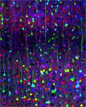 We may call this part of
the brain gray matter; but it doesnt look gray in these images.
These cortical neurons are involved in higher-thought processes
and perception of different senses.Although scientists have an idea of
the way neurons send messages to each other, there are still
many pieces of the puzzle that we dont know. For example, we still dont
know which neurons send messages to other neurons, where those neurons are located in the brain, or exactly which neurons receive messages from other neurons. Finding some order amongst the tangled chaos in the brain has been the goal of Dr. Jeff Lichtman and his laboratory at Harvard
University in Cambridge, MA. Over many years they have developed a technique to color-code different neurons and really light up your brain.
We may call this part of
the brain gray matter; but it doesnt look gray in these images.
These cortical neurons are involved in higher-thought processes
and perception of different senses.Although scientists have an idea of
the way neurons send messages to each other, there are still
many pieces of the puzzle that we dont know. For example, we still dont
know which neurons send messages to other neurons, where those neurons are located in the brain, or exactly which neurons receive messages from other neurons. Finding some order amongst the tangled chaos in the brain has been the goal of Dr. Jeff Lichtman and his laboratory at Harvard
University in Cambridge, MA. Over many years they have developed a technique to color-code different neurons and really light up your brain.
Jeff and his team of researchers are not
the first people to try to identify individual neurons. There
are several techniques that have been developed while attempting
to do the same thing. Each technique has encountered significant
difficulty that arises from the fact that the wires and cells
are not very far apart from each other. In fact some of them
are so close less than 200 nanometers (=
one hundredth the thickness of a hair) that even with a powerful
light microscope they cannot be seen as separate wires. Previous
techniques have used one, two, or three colors to differentiate
between different neurons and their axons and dendrites, but
that has only has limited capabilities. The technique developed
in the Lichtman lab is unique because it allows for the neurons
to appear in about 100 distinct colors. This technique creates
a multicolored or rainbow brain and is fondly called Brainbow.
Living Color
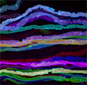
 Neurons in the auditory portion of the brain stem
On your computer or television screen you are able to see thousands
of colors of all different shades: yellows and oranges and purples
and blues. The screen is made up of many pinpoints of light,
called pixels.
Each pixel gives off a distinct color. But there arent thousands
of different pixel colors. In fact, your screen uses only three
colors of light red, green, and blue, or RGB that combine
in different amounts to get all the different colors you see.
In the same way that three colors of light yield the thousands
of shades you see on the screen, Brainbow uses three colors to
get about 100 different colors of distinct neurons.
Neurons in the auditory portion of the brain stem
On your computer or television screen you are able to see thousands
of colors of all different shades: yellows and oranges and purples
and blues. The screen is made up of many pinpoints of light,
called pixels.
Each pixel gives off a distinct color. But there arent thousands
of different pixel colors. In fact, your screen uses only three
colors of light red, green, and blue, or RGB that combine
in different amounts to get all the different colors you see.
In the same way that three colors of light yield the thousands
of shades you see on the screen, Brainbow uses three colors to
get about 100 different colors of distinct neurons.
The human body is much more complicated
than a computer or television screen, though. A screen uses multiple
colored pixels that show images: to get a similar variation in
the brain, you must use genes to make the neurons fluoresce,
or glow. It is not normally the case that neurons in the brain
give off various bright colors. The genes to enable the neurons
to do this must be inserted into the genetic material of the
organism whose brain is being looked at.
 Neurons in the hippocampus To find the genes that
enable organisms to glow (since mice do not generally fluoresce),
the Harvard group took advantage of some sea animals that do
give off light. They are known as bioluminescent organisms. In
particular they looked at jellyfish and types of corals that
glow naturally. When there is a certain level of calcium ions
in the cells of certain species of jellyfish, the jellyfish emit
blue light. (The jellyfish actually glow green, however, because
they have a gene that creates a protein that absorbs the blue
light and emits green light instead.) Jeff and his research colleagues
used three different genes that create three different proteins:
one that emits red light, one that emits green light, and one
that emits blue light. When these three genes are combined, they
can then be inserted into the genetic material of a mouse.
Neurons in the hippocampus To find the genes that
enable organisms to glow (since mice do not generally fluoresce),
the Harvard group took advantage of some sea animals that do
give off light. They are known as bioluminescent organisms. In
particular they looked at jellyfish and types of corals that
glow naturally. When there is a certain level of calcium ions
in the cells of certain species of jellyfish, the jellyfish emit
blue light. (The jellyfish actually glow green, however, because
they have a gene that creates a protein that absorbs the blue
light and emits green light instead.) Jeff and his research colleagues
used three different genes that create three different proteins:
one that emits red light, one that emits green light, and one
that emits blue light. When these three genes are combined, they
can then be inserted into the genetic material of a mouse.
The
technique of inserting foreign genes into the genetic material
of an organism is known as the transgenic method. In a fine and
complicated process, the Harvard team inserted the new genes
into mice, creating transgenic mice. All three genes are inserted
together in a microscopic cassette, and this cassette is inserted
into the genome in multiple copies. This technique allows for
the red, green, and blue colors to combine in each neuron in
a different way, yielding about 100 different colors of neurons in the brain.
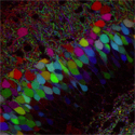
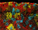
 Neurons in the dentate gyrus, part of the hippocampusSince the color of a single neuron is distinct
from the surrounding neurons, it is possible to follow the dendrites
and axons of one single neuron throughout the brain. In this
way it is also possible to see where the different wires in the
brain lead and how different neurons are related.
Neurons in the dentate gyrus, part of the hippocampusSince the color of a single neuron is distinct
from the surrounding neurons, it is possible to follow the dendrites
and axons of one single neuron throughout the brain. In this
way it is also possible to see where the different wires in the
brain lead and how different neurons are related.
How Our Brains Develop
As you can
imagine, there are many possible applications for the Brainbow
technique. Jeffs laboratory is primarily interested in observing the normal development of the brain. By observing the behavior of infants, toddlers, teenagers, adults, and seniors, we know that the human brain functions differently depending on age. The researchers hope to begin to understand some of these differences by directly observing changes in the brains wiring as animals grow up and grow old.
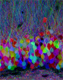
 Motor neuron axons from a portion of the oculomotor nerveWe know that many processes are happening
that allow children to learn to walk, talk, read, write, work,
etc. In a somewhat counterintuitive manner, you do not actually
create more brain cells as you learn. Instead, as you learn more
things, neurons in your brain make new connections, sorting through
the jumble of brain cells that already exist. The wires that
are not used may disappear or become inactive. For this reason,
if a child does not learn language before a certain age, that
child will never have normal language. If you try to learn a
second language after a certain age, it is very difficult to
speak with a native accent because the neurons that helped you
the first time are no longer in use.
Motor neuron axons from a portion of the oculomotor nerveWe know that many processes are happening
that allow children to learn to walk, talk, read, write, work,
etc. In a somewhat counterintuitive manner, you do not actually
create more brain cells as you learn. Instead, as you learn more
things, neurons in your brain make new connections, sorting through
the jumble of brain cells that already exist. The wires that
are not used may disappear or become inactive. For this reason,
if a child does not learn language before a certain age, that
child will never have normal language. If you try to learn a
second language after a certain age, it is very difficult to
speak with a native accent because the neurons that helped you
the first time are no longer in use.
These are the sorts of processes
that this group of neurobiologists plans to study. Of course,
this technology is not available in humans because it would involve
inserting new genetic information into an embryo. Humans also
develop over decades, which is not a practical time frame for
observation and research. The Lichtman lab instead is looking
at the brains of mice. Mice age over a period of weeks and provide
a good model for studying the mammalian brain. The researchers
use laser microscopes to study the colorful neurons in the brain
and how they change with age from birth, to maturity and old age.
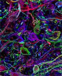
 Glial cells called astrocytes play a role in brain repairIn addition, they are currently collaborating
with other researchers and experimenting with other techniques
to improve upon Brainbow with more colors and better resolution.
They hope that improved techniques will enable them to study
structures in the brain that are too small to color-code.
Glial cells called astrocytes play a role in brain repairIn addition, they are currently collaborating
with other researchers and experimenting with other techniques
to improve upon Brainbow with more colors and better resolution.
They hope that improved techniques will enable them to study
structures in the brain that are too small to color-code.
Dr.
Jeff Lichtman is a Professor of Molecular and Cellular Biology
at Harvard University in Cambridge, Massachusetts. He was interested
in science from an early age, and he had a microscope in his
room since middle school. He attended Bowdoin College in Maine
where he majored in biology, and continued his education with
a Medical degree and Ph.D. in neuroscience.
In his free time he likes to play the piano and tend his garden.
To Learn More:
- Lichtman, J., J. Livet, and J. Sanes. A technicolor
approach to the connectome.
Nature Reviews 9 (2008): 417-422.
- Livet, J., T. Weissman, H. Kang, R. Draft, J. Lu, et
al. Transgenic strategies
for combinatorial expression of fluorescent proteins in the nervous system.
Nature 450 (2007): 56-62.
For More Information:
Written by Rebecca Kranz with Andrea
Gwosdow, Ph.D. Gwosdow
Associates
All photos (c) Livet, Weissman Draft, Sanes and Lichtman, Harvard University.
HOME | ABOUT | ARCHIVES | TEACHERS | LINKS | CONTACT
All content on this site is © Massachusetts
Society for Medical Research or others. Please read our copyright
statement — it is important. |
|
|
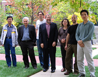
Co-authors and research colleagues on a recent paper in Nature on the mapping of neuronal circuits in Brainbow mice: (left to right): Ju Lu, Jeff W. Lichtman, Jean Livet, Joshua R. Sanes, Tamily A. Weissman, Ryan W. Draft, Hyuno Kang
Click
here to hear an interview with Jeff Lichtman on National Public Radio
Sign Up for our Monthly Announcement!
...or  subscribe to all of our stories! subscribe to all of our stories!

What A Year! is a project of the Massachusetts
Society for Medical Research.
|
|
