10/11 - How Strong Are Your Circuits? - MSMR: What A Year!
10.11 >> How Strong Are Your Circuits?
How Strong Are Your Circuits?
It's happened to everyone. You're taking a test, and you're doing fine, and then you freeze up. You didn't study for that question. Suddenly you've lost momentum and things start going downhill from there. We can't help you with the answers to those unexpected questions, but new findings by Dr. Amy Arnsten and her team of researchers at Yale University School of Medicine help explain why you stop doing so well. Perhaps after this story, the next time a test question catches you off guard, you'll be able to keep your cool.
Brain Basics
Let's begin with some brain basics. You may have learned about three main parts of the brain: the
cerebrum,
cerebellum, and
brain stem.
The brain stem controls our most basic functions necessary for survival, like our breathing, heart rate, and reflexes; the cerebellum plays an important role in functions like language, balance, and motor coordination. The cerebrum is the largest and most developed part of the brain involved in a variety of functions including thought, planning, and sensory perception. In fact, most of these functions occur in the outer layer of the cerebrum known as the
cerebral cortex.
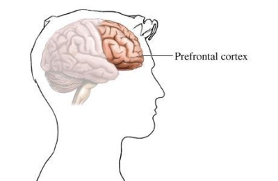
When you're studying for an exam, the cerebral cortex is the part of the brain that's most involved, particularly the front region known as the
prefrontal cortex.
The prefrontal cortex allows us to organize, plan ahead, and make good judgments. It is responsible for what's called
working memory
, the ability to retain what has just happened or bring to mind something from the past and use it to guide our future thoughts and actions. Working memory provides the foundation for the formation of abstract thoughts.
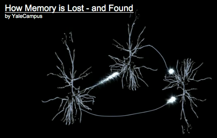
All brain function depends on the specialized cells called
neurons
that have the ability to communicate with other neurons using electrochemical signals. Neurons come in all shapes and sizes, but they all have the same basic parts:
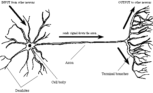
The
dendrites
of the cell body are extensions that receive messages from other neurons and transmit them back to the cell body. There can also be specialized protrusions on the dendrites called spines that often receive messages from other neurons in the cerebral cortex.
The
axon
of a cell body is an extension that transmits messages to other neurons when excited by the cell body.
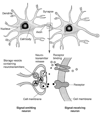
Most neurons have a cell body with many dendrites and an axon that connects with a large number of other neurons. The axon communicates with other neurons across a very small space called the
synapse.
We said earlier that neurons use electrochemical signals to transmit messages across the network. What makes neurons so special is their ability to conduct electricity—that's the "electro" part. When the dendrite of a neuron receives a message, it sends an electrical current to the cell body, which sends that same current out to the axon. The electrical current is created by the passage of positively and negatively charged molecules called
ions
in and out of the cell. The electrical impulse is carried down the axon in what is called an
action potential
when a neuron does this we say that the neuron is "firing". The electrical signal can't cross the synapse at the end of the axon, though, so here the electrical signal is converted into a chemical signal—the "chemical" part. The electrical impulse stimulates the axon to send chemicals called
neurotransmitters
across the synapse. You may be familiar with some neurotransmitters like
epinepherine
, (formerly known as adrenaline), the active ingredient in epi-pens. Neurotransmitters are received by specialized proteins called receptors that that recognize and bind to specific neurotransmitters like a key fitting into a lock. When the lock opens, charged compounds called ions come into the neuron, creating an electrical impulse that can go to the cell body and on to the axon. If the signal is excitatory, it will be passed on to the next neuron. If not, the signal is said to be inhibitory, and it won't be passed to other neurons.
In the past several decades,
neurobiologists
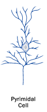
(scientists who study the brain) have made great strides in understanding how our neurons work together to form thoughts and execute actions. Researchers have known for many years that neurons fire in response to environmental cues, even something as basic as your eye movements while you're reading this article. It was only in recent years that they have come to understand that working memory depends on the continual firing of neurons in the prefrontal cortex, even in the absence of external stimuli. These neurons, known as
pyramidal cells
for their triangular-shaped cell body, make up the majority of excitatory neurons in the prefrontal cortex. The pyramidal cells are arranged in circuits–that excite each other to maintain neuronal firing and keep information "in mind". These circuits are very sensitive to the many neurotransmitters and other compounds that surround them. In fact, new research by Dr. Arnsten and her team of scientists at Yale Medical School has shown that relatively subtle chemical changes that occur even with mild stress can alter the firing of prefrontal pyramidal cells.
Their research shows that when you're under stress–even very mild stress like an unexpected exam question–the class of neurotransmitters that epinephrine belongs to, called catecholamines, are released in your prefrontal cortex. The
catecholamines
cause ion channels on dendritic spines to open where they would normally be closed. In the same way that holes in a garden hose prevent all the water from reaching your flowers, these opened ion channels weaken the ability of pyramidal cells to communicate with each other. Since the pyramidal cells can no longer excite each other to keep information "in mind", your working memory becomes impaired. As a result, it becomes more difficult to recall what you spent several hours studying the evening before. This weakening of connections between neurons also occurs naturally as part of the aging process. This is why your parents and grandparents have more difficulty remembering where they put things or recalling someone's name.
Dr. Arnsten began this research by observing the functioning of individual neurons in the brains of monkeys as they performed working memory tasks. "We've had these monkeys for over twenty years," Dr. Arnsten explained. "They're really our partners in this research." The tasks are performed on a computer like a video game, and the monkeys set the pace for themselves. The computer is equipped with a machine that can track where the monkeys' pupils are. When the monkey stares at the "X" in the middle of the screen, the researchers know it's ready to begin. At the start of the trial, a light will flash randomly in one of eight locations around the center of the screen. There will then be a delay of between 2.5 and 5 seconds during which the monkey is supposed to remember where the light flashed. After the allotted number of seconds, the "X" will disappear from the center of the screen, and the monkey can move its eyes to the remembered location to receive some juice as a reward. The next trial will begin when the monkey is ready and staring at the "X" in the center. [If this test were done with humans, the subjects could 'cheat' by repeating, using language, what they were supposed to remember. Monkeys don't have language, so this is an appropriate working memory task.]
Dr. Arnsten found that older monkeys did not perform as well and rested more frequently than younger monkeys. When she looked at the neuronal function of the monkeys as they performed the task, she was able to hypothesize why this might be. The older monkeys actually had weaker neuronal activity than the younger monkeys during the delay when they were supposed to remember where the light had flashed.
Next Step: Tiny Gold Balls
Next, Dr. Arnsten wanted to understand the chemistry behind the observed weakening of the connections between pyramidal cells. To do this, she used a technique known as
immunoelectromicroscopy
. Immunoelectromicroscopy takes advantage of gold balls that come in many different sizes and can be seen using an electron microscope, which can see things that would be invisible to your ordinary light microscope. The gold balls are attached to molecules that recognize and bind to specific proteins in the brain; the gold balls thus serve as a marker for that protein. In the resulting image, researchers can see where the different molecules are located by the size and location of the gold balls. When Dr. Arnsten and her team viewed the resulting images, they saw that signaling molecules were clustered together in spines right near the synapse. These results, combined with the previous findings in her work with monkeys, led Dr. Arnsten to the idea that the increase in signaling molecules near the synapse resulted in the weakened connections observed as cognitive defects associated with aging and stress.
Some Good News
Fortunately, the weakening of connections between neurons appears to be reversible. When Dr. Arnsten and her team inhibited the signaling molecules or blocked the ion channels, they found that the connections did indeed become more robust. A variety of compounds were tested in these experiments. One of these compounds, called
guanfacine
, is approved by the Food and Drug Administration for treating attention deficit disorders in children and high blood pressure in adults. Yale Medical School is currently conducting trials in humans, known as
clinical trials
, to determine whether guanfacine can reverse some of the cognitive defects associated with aging and stress in humans.
Dr. Arnsten's ultimate goal is to help people overcome the cognitive deficits associated with aging and mental illness. For example, there is some evidence that the right side of the prefrontal cortex is weaker in people with
Attention Deficit Hyperactivity Disorder (ADHD), which makes them easily distracted. Guanfacine helps people with ADHD to be less distracted and less impulsive. Similarly, studies have shown that guanfacine may help people to quit smoking even under stress by strengthening their prefrontal cortex and allowing them to have more self-control.
A Better Understanding of the Brain
Dr. Arnsten is currently trying to understand what is happening in the brain at the molecular level. Dr. Arnsten hopes her work will reveal why older brains are vulnerable to degeneration and why the prefrontal cortex is vulnerable to mental illnesses like
schizophrenia
. The results from these human trials won't be available for at least several more years. In the meantime, keep studying for your exams. But the next time you get a curveball, maybe you'll be able to help your pyramidal connections stay just a bit stronger by relaxing, and letting those circuits reconnect.
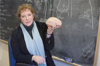
Dr. Amy Arnsten is a Professor of Neurobiology at Yale Medical School. She studies the molecular basis of cognitive functions of the prefrontal cortex with the goal of developing treatments for cognitive defects associated with aging and mental illness. Dr. Arnsten became interested in the brain and mental illness after observing how poorly people with mental illness are treated in our society. When not in the lab, Dr. Arnsten enjoys hiking and spending time with her family.
For More Information:
Arnsten, A. 2009. "Stress signaling pathways that impair prefrontal cortex structure and function." Nature Reviews Neuroscience, 10: 410-422.
Arnsten, A. et al. "Dynamic Network Connectivity: A new form of neuroplasticity." Trends in Cognitive Sciences, 14(8): 365-375.
Wang, M. et al. 2011. "Neuronal basis of age-related working memory decline."Nature, 476: 210-213.