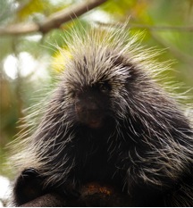
Those 30,000-plus quills will disengage when the animal feels threatened, and lodge themselves painfully and solidly into your skin. This is because the North American porcupine quill tips have tiny backward-facing barbs that allow the quills to easily penetrate tissue but make them very difficult to remove. While a face full of quills is certainly to be avoided, in the laboratory of Dr. Jeffrey Karp, they have become inspiration for surgical-grade needles and adhesives.
Porcupine
When Dr. Karp began investigating porcupine quills for biomedical applications, he was surprised to find that little previous research had been conducted describing quill biomechanics and function. That got us thinking, remarks Dr. Karp. We knew we might be on to something. He recognized that the backward-facing barbs on the tips of the quills were critical to their effectiveness.
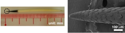
Natural porcupine quill: digital photo and scanning electron microscope
In order to quantify the contribution of the barb compared to the rest of the quill, Dr. Karp conducted a series of experiments on barbed quills and quills that the researchers had modified by removing the barbs. The researchers measured two forces: the force required to insert the quill, known as the penetration force; and the force required to extract the quill, known as the pull-out force. These experiments were performed using a machine that could uniformly insert the quills and precisely measure the forces. Similar trials were conducted with samples of muscle tissue,
porcine skin,
and synthetic human skin.
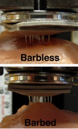
Synthetic quills with and without barbs
The results of these experiments were quite remarkable. The barbed quills of the North American porcupine required 54 percent less penetration force than the barbless quills, meaning that they entered the tissue much more easily. The penetration force of the barbed quills was also significantly less than that of hypodermic needles currently used for a variety of purposes in medical and surgical settings.
The pull-out force was much greater in the barbed quills compared to the barbless quills. The researchers confirmed their results by conducting the same experiments on both barbed North American porcupine quills and naturally unbarbed African porcupine quills. The researchers found similar findings, that the pull-out force was much less in the barbed quills.
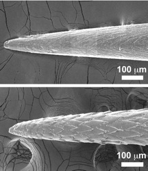
Tips of barbless (top), and quills with barbs-mimetic needles; scanning electron microscope images
These results prompted the researchers to create synthetic porcupine-inspired needles with microscopic backward-facing barbs at the tip. When tested against synthetic barbless needles, the researchers found that the barbed needles required 80 percent less penetration force. When the researchers wove the synthetic quills together into an interlocking adhesive pattern, they found that the barbed quill patch had significantly greater adhesive capacity than the barbless patch.
Video shows the differing adhesive qualities of barbed vs barbless quills
These findings have the potential for the development of new and better needles and adhesives that would penetrate the skin more easily and less painfully, while also remaining lodged in the tissue more solidly. Dr. Karp is pursuing this possibility through more experiments designed to optimize barb size, placement, and patterning.
This work with porcupine quills is only one example of the research conducted by Dr. Karp in his laboratory, which uses bioinspired biology and medical knowledge to solve medical problems. His multidisciplinary team includes biologists, engineers, clinicians, and medical students with diversified areas of expertise to pursue novel approaches that can be rapidly translated into solutions for clinical use.
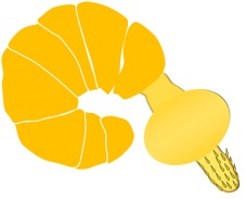
A cartoon parasite with a barbed proboscis
Dr. Karp is also working on another needle design inspired by the intestinal parasite Pomphorhynchus laevis, which attaches itself to the intestinal wall of fish by swelling a bulb at the tip of its
proboscis
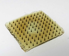
after penetration of the tissue. The researchers have taken this idea of a swellable tip into the laboratory, where they have developed an adhesive array of very small needles, called microneedles, with swellable tips.
Adhesive array of microneedles
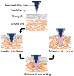
Preliminary tests of the design were conducted using pig skin, which is a good model for human skin (link to video: adhesive comparison.wmv). The results of these studies were promising, and further experiments are now ongoing in live animals. Dr. Karp and his team are hopeful that the swellable microneedles will prove to be a successful adhesive device that helps reduce infection and accelerate tissue regeneration. Here's how they work:
Swellable tip mechanism
Sutures and Adhesive Devices
Dr. Karp and his team are involved in several projects to improve suture and adhesive devices, a field that has not seen much innovation in recent years. Adhesive devices are tricky, because they must remain in place for a very specific amount of time but must also be easily placed, and sometimes also easily removed, depending on the type of device. Available options include chemical adhesives, sutures, and staples, all of which can potentially result in complications. Chemical adhesives are often highly reactive and toxic, and may stimulate a strong inflammatory response as they degrade. Sutures require realignment of the tissue with each pass of the suture needle, which can be very time consuming and increases the risk for complications and infection. Staples are quicker, but they create larger holes that can become sites for bacterial infiltration and infection, and they can damage tissue both on the way in and the way out.
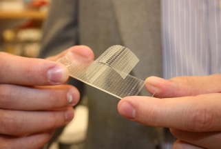
Dr. Karp and his team are in search of bioinspired adhesive devices, like the porcupine-inspired needles described above. Currently, a specific problem is tearing of thin skin, which is found on premature babies and in the elderly. Dr. Karp solved this problem by inserting a third middle layer into the tape, which decouples the application of the tape from its removal.
Karp Quick Release Medical Tape
He started by testing his model on origami paper, which is very frail and tears easily. He measured the forces of adhesion and the point of breakage. When his design worked on origami paper, he tested the tape on pigskin. He eventually hopes to bring his design to the clinic, which is one of the goals of his research.
Quick-release Medical Tape Demonstration
Dr. Jeffrey Karp is Co-Director of the Center for Regenerative Therapeutics at Brigham and Womens Hospital in Boston and an Associate Professor at Harvard Medical School. His laboratory includes a multidisciplinary team of researchers, clinicians, and students with five areas of focus: tissue adhesives, cell therapy, drug delivery, diagnostic devices, and medical devices. In addition to developing new technologies and therapies for clinical use, Dr. Karp also hopes to use his multidisciplinary, collaborative approach to change the way we approach scientific problems and train the next generation of bioengineers. Twelve post-doctoral fellows from Dr. Karps lab have already transitioned into faculty positions. When not in the laboratory, Dr. Karp enjoys walking several miles each way to work and spending time with his family.
For More Information:
Yang, S. et al. 2013. A bio-inspired swellable microneedle adhesive for mechanical interlocking with tissue. Nature Communications, 2013;4:1702. doi: 10.1038/ncomms2715.
Cho, W. et al. 2012. Microstructured barbs on the North American porcupine quill enable easy tissue penetration and difficult removal. Proceedings of the National Academy of Sciences, 2012 Dec 26;109(52):21289-94. doi: 10.1073/pnas.1216441109. Epub 2012 Dec 10..
Zhao, W. et al. 2012. Bioinspired multivalent DNA network for capture and release of cells. Proceedings of the National Academy of Sciences, 2012 Nov 27;109(48):19626-31. doi: 10.1073/pnas.1211234109. Epub 2012 Nov 12.
Laulicht, B. et al. 2012. Quick-release medical tape. Proceedings of the National Academy of Sciences, 2012 Nov 13;109(46):18803-8. doi: 10.1073/pnas.1216071109. Epub 2012 Oct 29.
Mahdavi, A. et al. 2008. A biodegradable and biocompatible gecko-inspired tissue adhesive. Proceedings of the National Academy of Sciences, 2008 Feb 19;105(7):2307-12. doi: 10.1073/pnas.0712117105. Epub 2008 Feb 19.
Written by Rebecca Kranz with Andrea Gwosdow, PhD at www.gwosdow.com